Patient Information
Cataract, AMD (IVT), epiretinal membrane, macular hole: patient information by Dr Julien Gozlan (Paris 16).
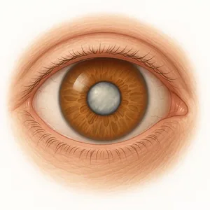
Cataract
Cataracts, phacoemulsification surgery, monofocal and multifocal lenses...
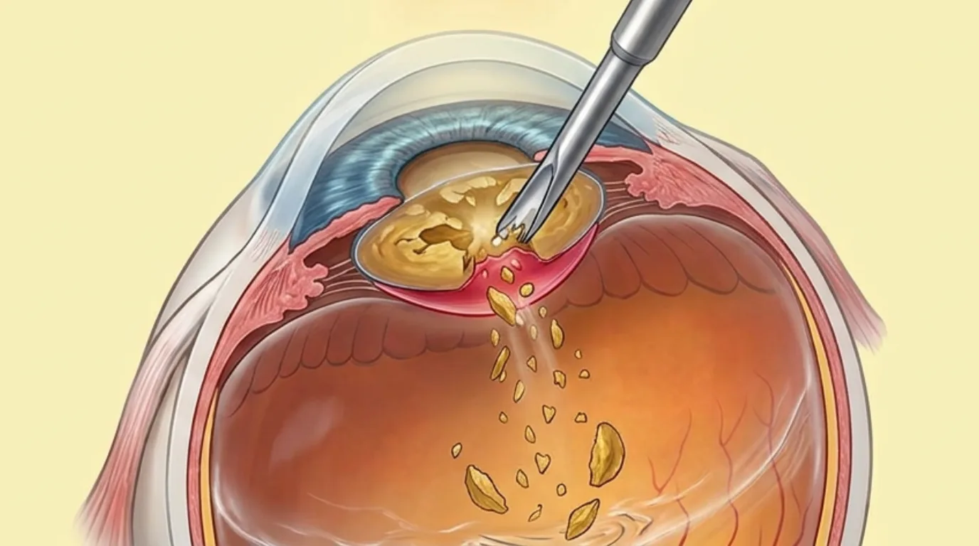
Posterior capsular rupture
Also covered onCataracte.fr

Cataract
Also covered onCataracte.fr
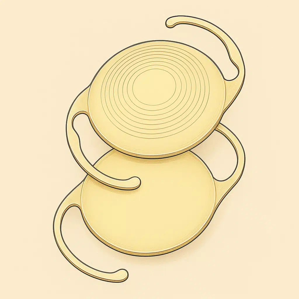
Cataract Lens Implants
Also covered onCataracte.fr
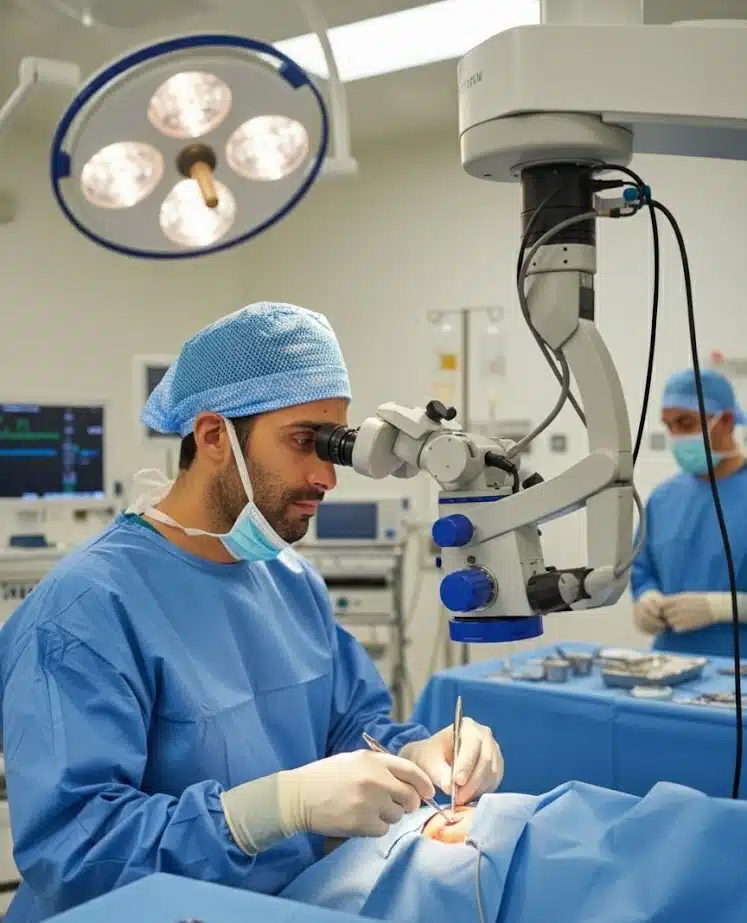
Cataract Surgery
Also covered onCataracte.fr
Cataract Surgery by a Retina Surgeon
Choosing a retina surgeon for your cataract surgery offers a considerable advantage in terms of safety and quality of care. Dr Julien Gozlan, an ophthalmologist surgeon in Paris 16, combines expertise in cataract surgery and vitreoretinal surgery. This dual competence allows him to manage all intraoperative situations, including the most complex ones, and to offer combined procedures when the retina requires simultaneous treatment. This article explains why entrusting your cataract to a retina surgeon provides additional peace of mind, from complication management to the treatment of associated retinal conditions.
Read more

Femtosecond laser cataract surgery
Femtosecond laser cataract surgery raises many questions among patients considering lens surgery. Presented as a major technological advancement, the femtosecond laser applied to cataract surgery is the subject of intensive marketing, but the international scientific literature reveals specific limitations and risks compared to conventional manual phacoemulsification. Dr Julien Gozlan, ophthalmologist in Paris 16, explains why the manual technique remains the safest and most reproducible standard for cataract surgery.
Read more
Retinal Diseases
Age-related macular degeneration (AMD), intravitreal injections (IVT), diabetic retinopathy, Irvine-Gass syndrome...
Eye Floaters
Also covered onRetine.fr

Choroidal Metastasis
Choroidal metastasis refers to a secondary tumour localisation within the choroid, the vascular membrane situated between the retina and the sclera of the eye. Dr Julien Gozlan, ophthalmologist surgeon specialising in the retina in Paris 16, manages patients presenting with this ocular condition, which is often an indicator of a distant cancer. This article details the mechanisms of choroidal metastasis, the symptoms that should raise concern, the essential diagnostic examinations and current therapeutic options, presented in a reassuring and educational approach.
Read more
Choroidal Naevus
A choroidal naevus is a benign pigmented lesion located at the back of the eye, often discovered incidentally during a routine ophthalmological examination. Comparable to a skin mole, this naevus develops in the choroid, the vascular membrane that nourishes the retina. Dr Julien Gozlan, ophthalmologist surgeon specialising in the retina in Paris 16, explains in this article what a choroidal naevus is, how it is diagnosed, what the monitoring criteria are, and in which cases specific management is required.
Read more
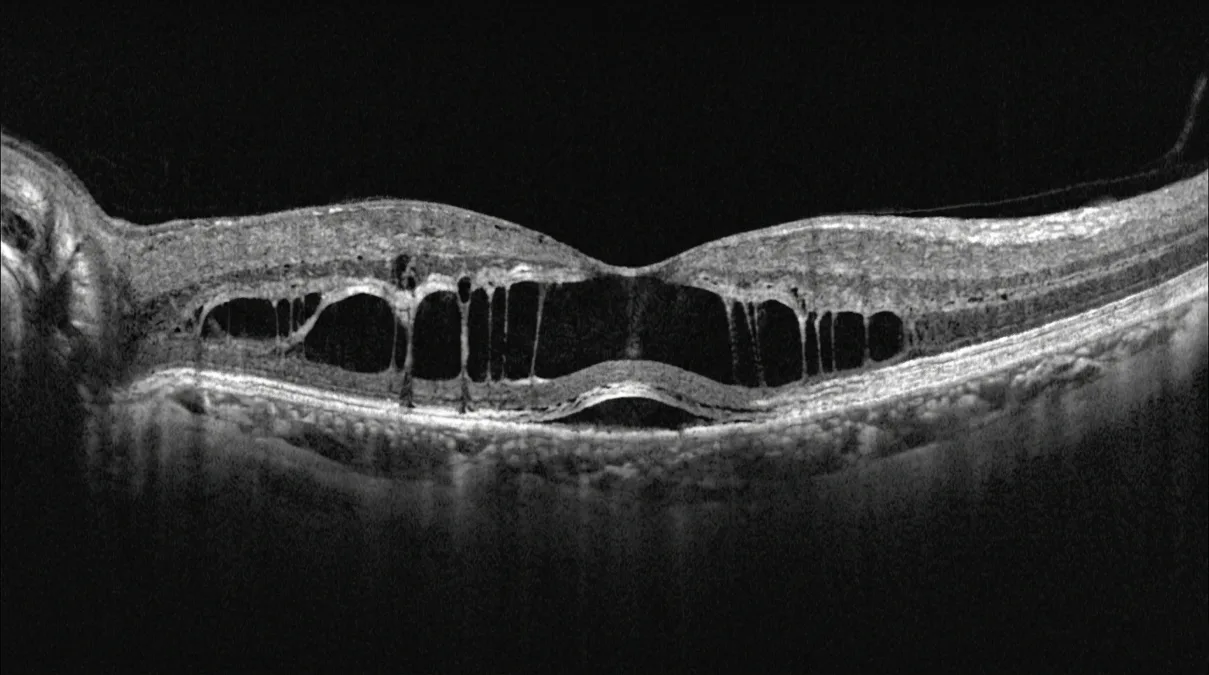
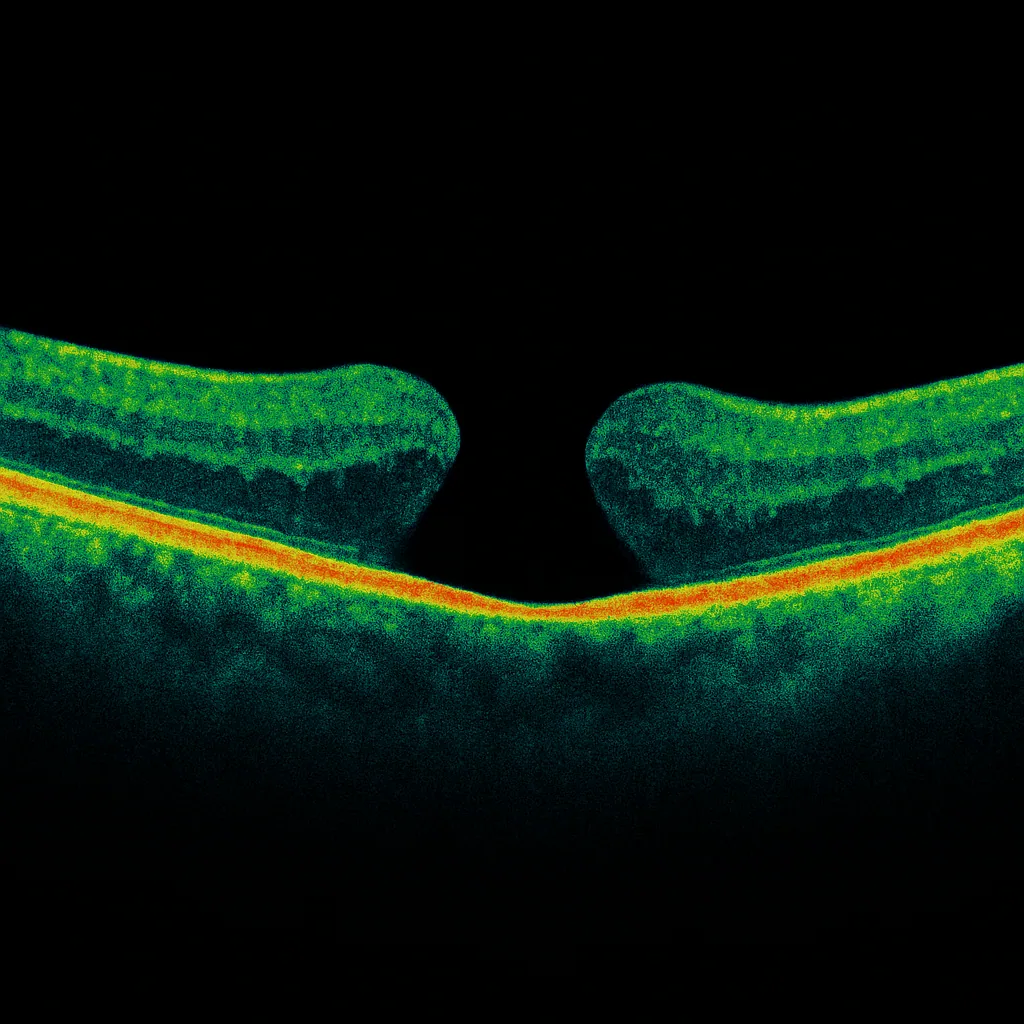
Myopic Foveoschisis
Myopic foveoschisis is a degenerative macular condition specific to high myopia, characterized by progressive splitting of the retinal layers at the fovea. Long underdiagnosed, this condition is now identified thanks to optical coherence tomography (OCT). Dr Julien Gozlan, ophthalmic surgeon specializing in vitreoretinal surgery at Cabinet Ophtalmologique Paris – Auteuil, offers a comprehensive overview of this disease: definition, pathophysiological mechanisms, symptoms, imaging diagnosis, surgical indications, operative techniques, and visual prognosis.
Read more
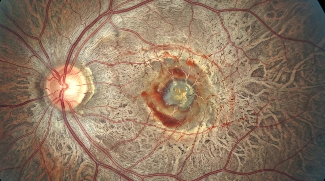
Choroidal neovascularization in high myopia
Choroidal neovascularization in high myopia is one of the most feared ocular complications in highly myopic patients. Dr Julien Gozlan, an ophthalmologist and surgeon specializing in retinal diseases at Cabinet Ophtalmologique Paris – Auteuil, manages this macular condition using state-of-the-art diagnostic equipment including OCT and OCT angiography. This article details the pathophysiological mechanisms of myopic choroidal neovascularization, its symptoms, current diagnostic methods, treatment with intravitreal anti-VEGF injections, and long-term visual prognosis.
Read more
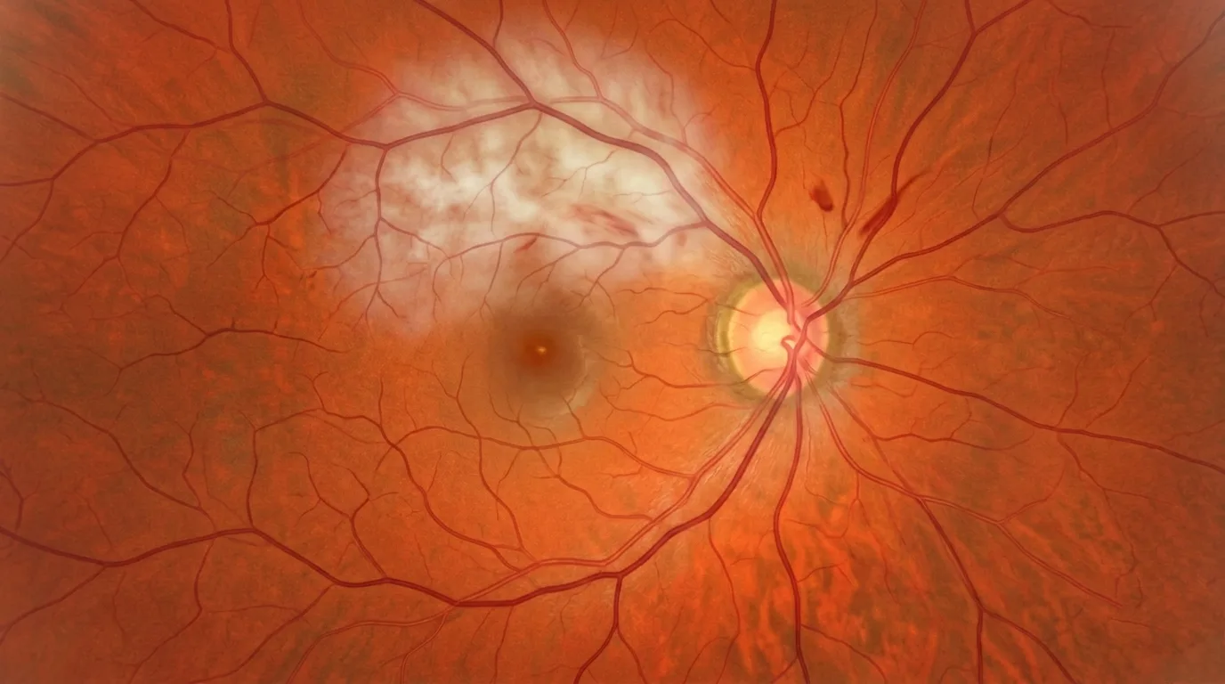
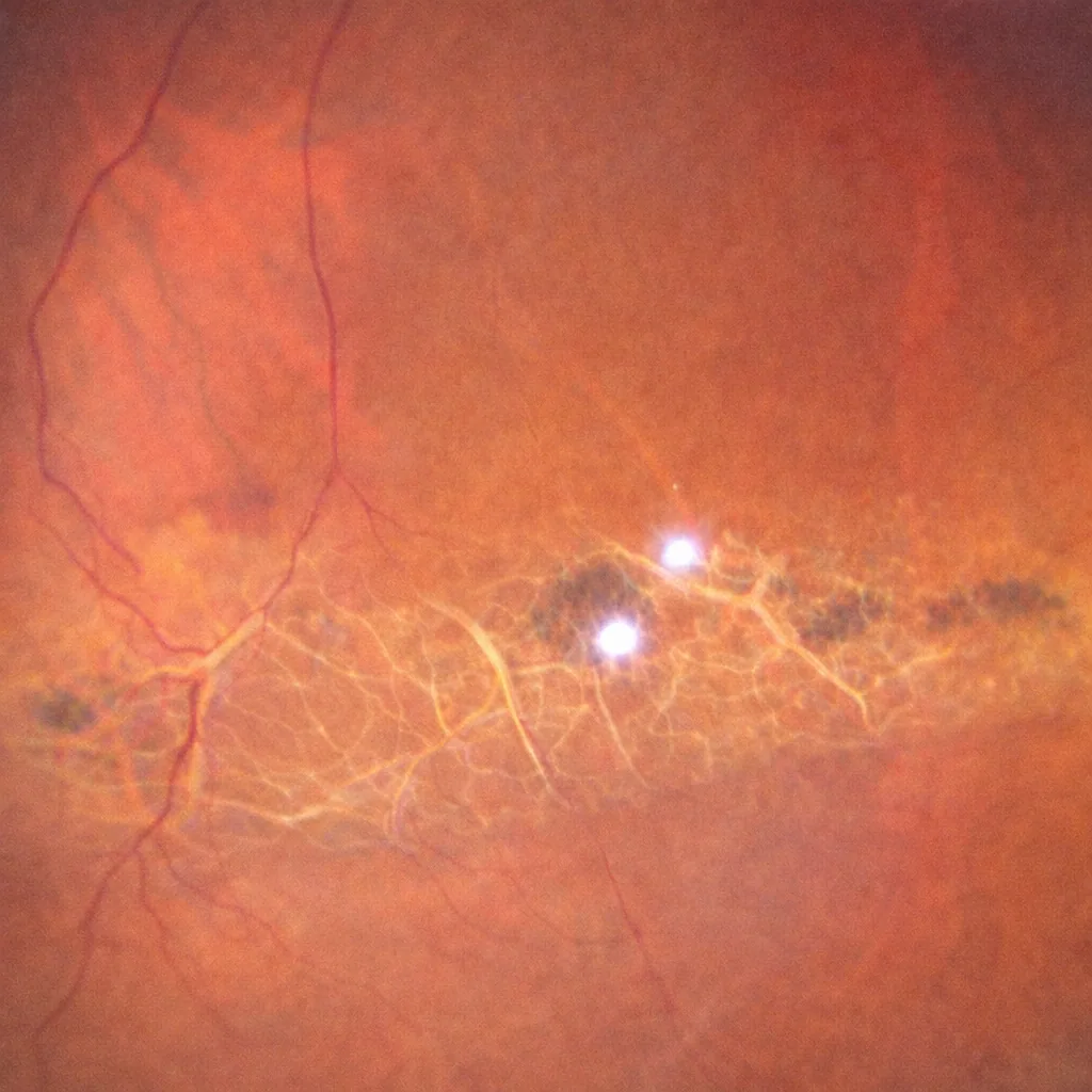
Retinal artery occlusion
Retinal artery occlusion is an ophthalmological emergency characterised by sudden loss of vision, comparable to a stroke of the eye. This rare but serious condition results from the interruption of blood flow in an artery supplying the retina, causing ischaemia that can lead to irreversible damage within hours. Dr Julien Gozlan, retina specialist at Cabinet Ophtalmologique Paris – Auteuil, explains in this article the different forms of retinal artery occlusion, their causes, warning symptoms, diagnostic examinations performed (fundus examination, OCT, OCT angiography), emergency management, visual prognosis and the critical importance of the associated cardiovascular workup.
Read more
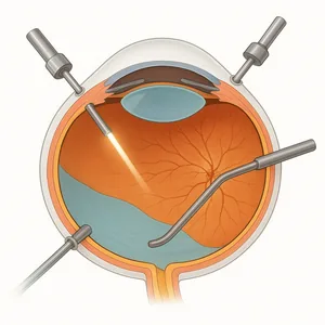
Retinal Surgery
Vitrectomy, retinal detachment, epiretinal membrane, macular hole...
Macular Epiretinal Membrane
Also covered onRetine.fr
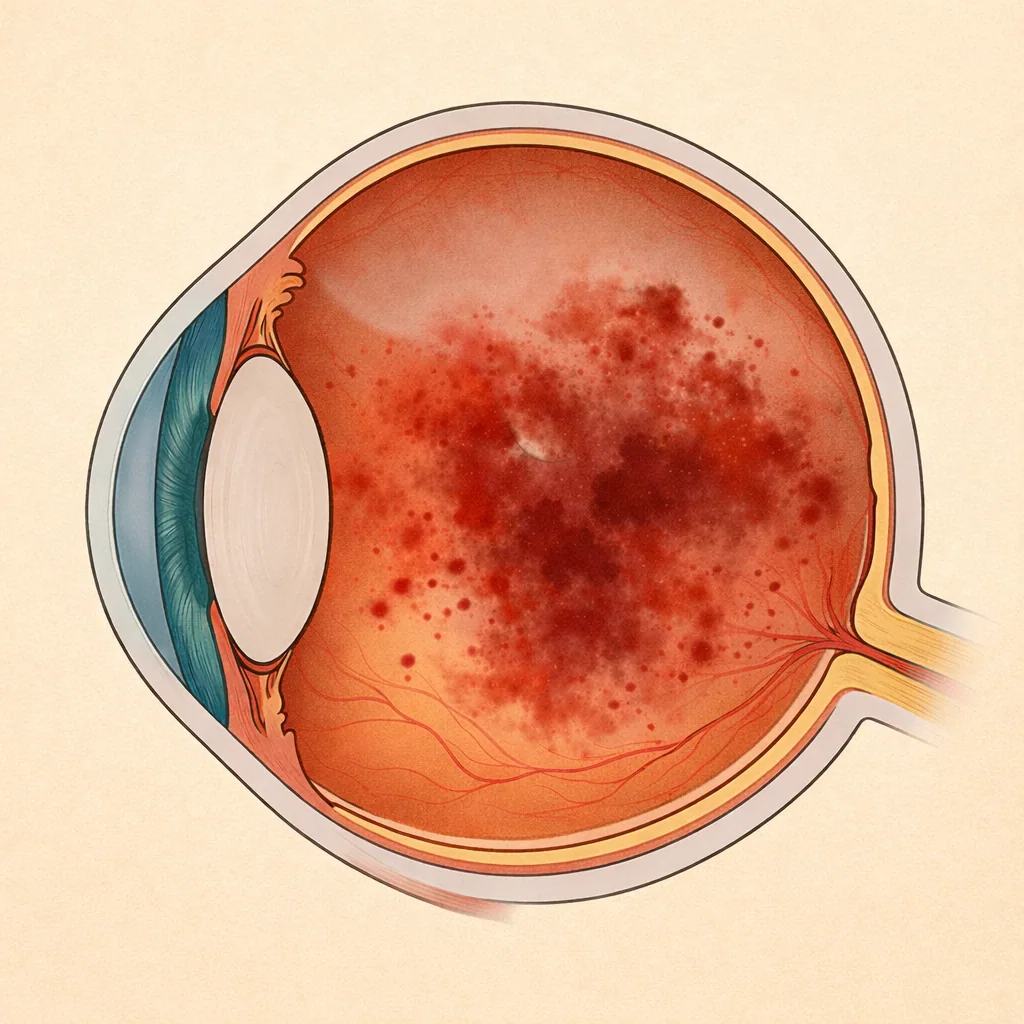
Vitreous Hemorrhage
Also covered onRetine.fr
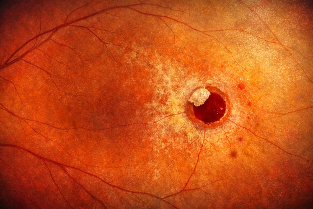
Macular Hole
Also covered onRetine.fr
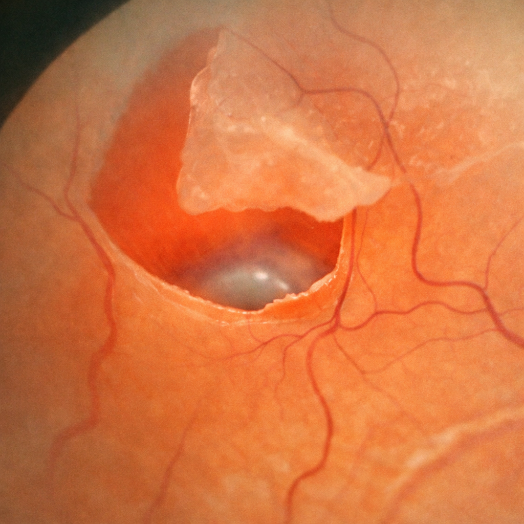
Operculated retinal hole
An operculated hole is a lesion of the peripheral retina caused by a posterior vitreous detachment (PVD). During this physiological process, the vitreous exerts traction on the retina and tears away a small fragment of retinal tissue, called an operculum, which remains suspended above the hole. This lesion is managed either through simple monitoring or preventive laser retinopexy performed in the office; surgery is only indicated in cases of associated retinal detachment. Dr Julien Gozlan, ophthalmologist in Paris 16, explains in this article the mechanisms of formation, symptoms, diagnosis, and management of this peripheral retinal lesion.
Read more
Lattice Degeneration
Lattice degeneration, also known as lattice retinal degeneration, refers to thin, weakened areas of the peripheral retina, often discovered during a routine fundus examination. It is generally asymptomatic but can increase the risk of retinal tears and subsequent retinal detachment in certain specific situations. Dr Julien Gozlan, ophthalmologist in Paris 16, explains what lattice degeneration is, when it should simply be monitored, and in which cases prophylactic laser retinopexy is truly indicated.
Read more
Retinal tear
A retinal tear is a break in the retinal tissue, often caused by traction from the vitreous (the gel that fills the eye). It can remain silent for a long time or present with floaters, flashes of light, or a curtain effect in the visual field. The main risk is progression to a retinal detachment, which constitutes a surgical emergency. Dr Julien Gozlan, ophthalmologist in Paris 16, explains the warning symptoms, diagnosis, laser treatments, and the follow-up that should be implemented.
Read more
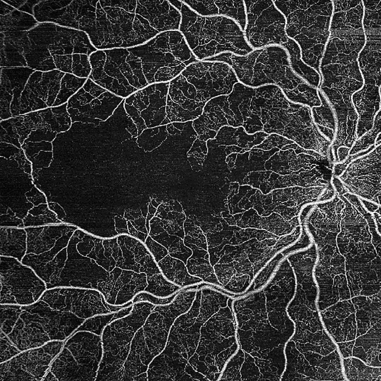
OCT Angiography
OCT angiography (or OCT-A) is a recent imaging technique that allows visualization of blood circulation in the retina and choroid without dye injection. It complements standard macular OCT and fluorescein angiography. Dr Julien Gozlan, ophthalmic surgeon in Paris 16, explains the principle, indications, and limitations of OCT angiography.
Read more
Indocyanine Green Angiography
Indocyanine green angiography (ICG) is an imaging examination that highlights the choroidal circulation, located beneath the retina. Complementary to fluorescein angiography and macular OCT, indocyanine green angiography aids in diagnosing choroidal conditions and guides their treatment.
Read more

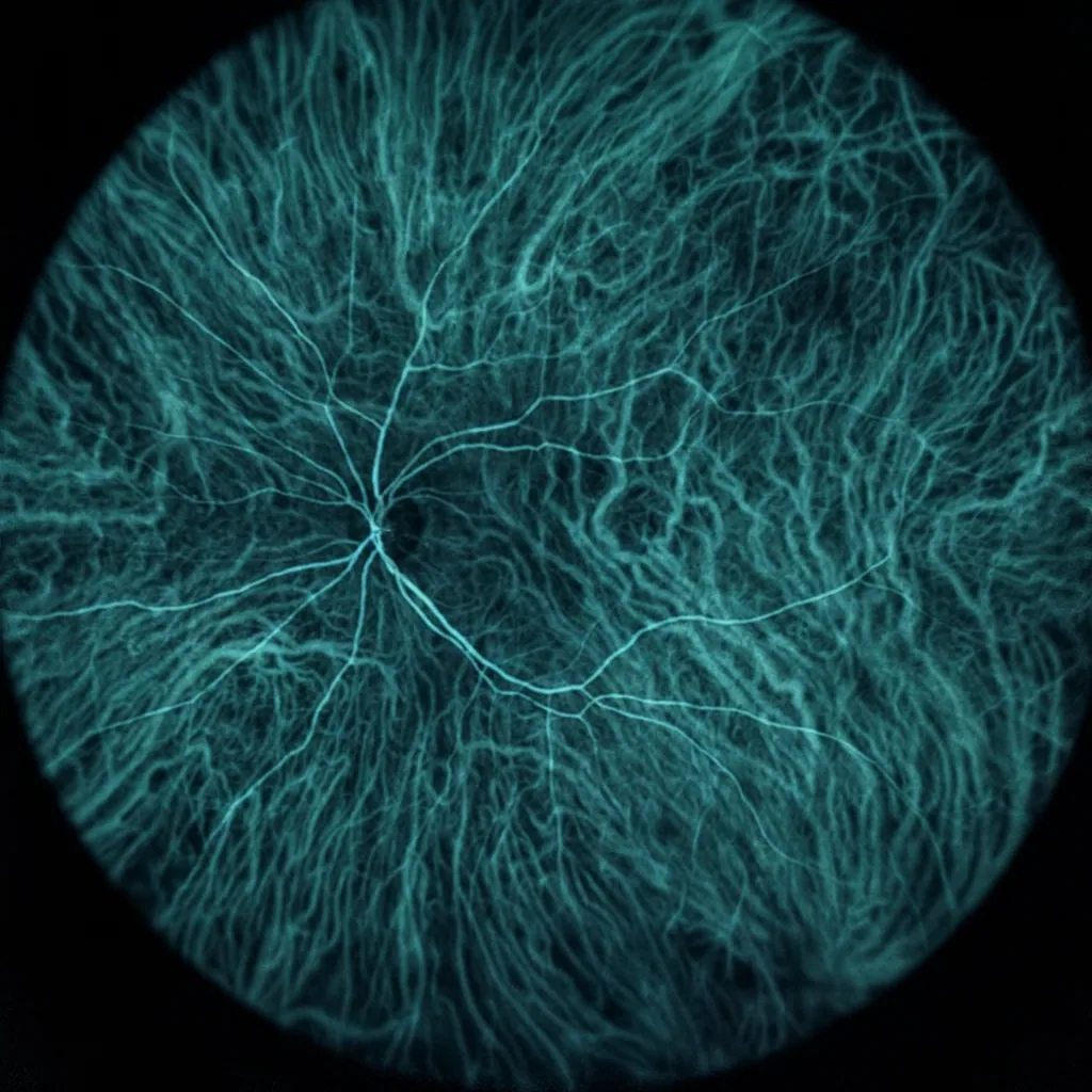
Fluorescein Angiography
Fluorescein angiography is a retinal imaging examination that allows observation of blood circulation and identification of leaks, hemorrhages, or ischemia. Performed in the office, this examination helps establish a precise diagnosis and guide the treatment of macular conditions such as AMD, or retinal vascular disorders such as CRVO. Dr Julien Gozlan explains its principle, indications, procedure, and risks.
Read more
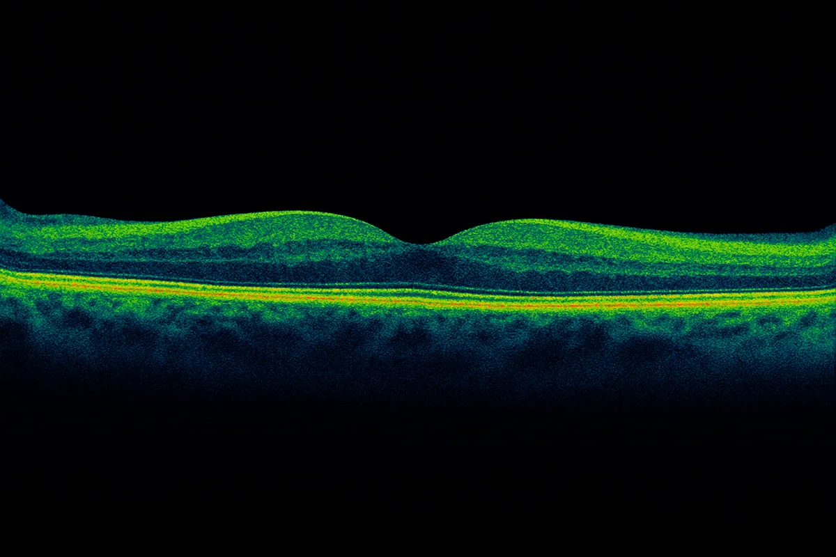
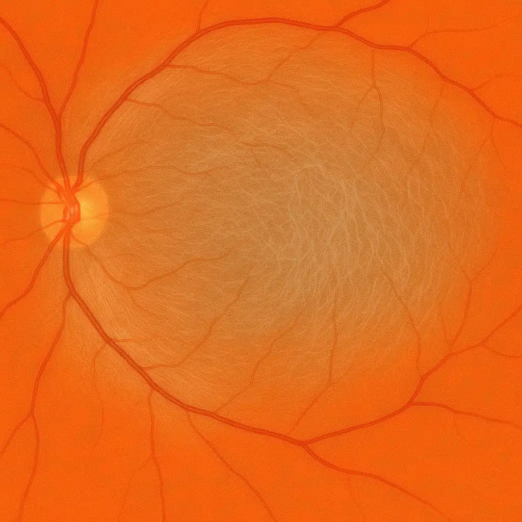
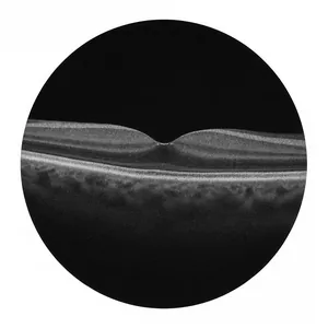
Optical Coherence Tomography
OCT (optical coherence tomography) is a retinal imaging exam that has become essential in ophthalmology. It allows highly precise analysis of the macula – the central area of the retina responsible for fine vision. Dr Julien Gozlan, ophthalmic surgeon in Paris 16, explains what macular OCT involves, when it is indicated, and how the examination is performed.
Read more