
A choroidal naevus is a benign pigmented lesion located at the back of the eye, often discovered incidentally during a routine ophthalmological examination. Comparable to a skin mole, this naevus develops in the choroid, the vascular membrane that nourishes the retina. Dr Julien Gozlan, ophthalmic surgeon specialising in the retina in Paris 16, explains in this article what a choroidal naevus is, how it is diagnosed, what the monitoring criteria are, and in which cases specific management is necessary.
What is a choroidal naevus?
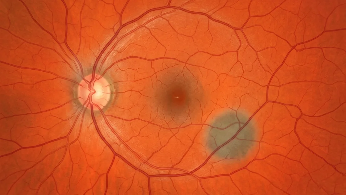
A choroidal naevus corresponds to an accumulation of melanocytes — the cells responsible for pigmentation — in the choroid. This vascular layer, located beneath the retina, supplies oxygen and nutrients to the photoreceptors. A choroidal naevus appears as a flat, greyish or brownish spot, generally small in size (less than 5 mm in diameter and less than 2 mm thick).
It is the most common benign intraocular tumour in adults: it is estimated that 5 to 10% of the population has a choroidal naevus, often without knowing it. In the vast majority of cases, this lesion remains stable and causes no visual symptoms. It is most often discovered during an fundus examination or an OCT performed for another reason.
Causes and risk factors of choroidal naevus
The exact causes of the appearance of a choroidal naevus are not fully understood. As with skin moles, several factors are involved:
- Genetic predisposition: individuals with fair skin and light eyes have a slightly higher incidence.
- Ultraviolet exposure: although the link is less direct than for cutaneous naevi, cumulative sun exposure may play a role.
- Age: choroidal naevus is more frequently diagnosed from the age of 50 onwards, possibly because fundus examinations become more regular.
It is important to emphasise that a choroidal naevus is not linked to a retinal disease such as age-related macular degeneration (AMD) or diabetic retinopathy. It is a distinct entity of melanocytic origin.
Symptoms and diagnosis of choroidal naevus
Is a choroidal naevus symptomatic?
In the vast majority of cases, a choroidal naevus is entirely asymptomatic. The patient experiences neither pain nor loss of vision. When located in the peripheral retina, it does not affect visual function in any way. Rarely, a naevus located near the macula may cause slight image distortion (metamorphopsia) or a mild visual haze, due to an associated accumulation of subretinal fluid.
Essential additional examinations
Diagnosis is based on a thorough fundus examination, supplemented by high-precision retinal imaging:
- OCT (optical coherence tomography): this non-invasive examination allows the thickness of the choroidal naevus to be measured and any associated abnormalities to be detected (subretinal fluid, retinal pigment epithelium alterations).
- OCT angiography: this visualises the vasculature of the naevus without dye injection.
- Fundus autofluorescence: this highlights the presence of lipofuscin (orange pigment) on the surface of the naevus, a sign of metabolic activity to be monitored.
- Indocyanine green angiography: this assesses deep choroidal circulation.
- B-mode ocular ultrasound: this precisely measures the thickness of the lesion and its echogenicity.
These examinations allow the choroidal naevus to be precisely characterised and differentiated from an early choroidal melanoma.
Choroidal naevus and risk of transformation: monitoring criteria
The main concern with a choroidal naevus is the — very low but real — risk of malignant transformation into a choroidal melanoma. This risk is estimated at approximately 1 in 8,000 per year for a typical naevus. To assess this risk, ophthalmologists use mnemonic criteria grouped under the acronym TFSOM-UHHD (or, in simplified form, the risk factors known as "MOLES"):
- T — Thickness: a naevus more than 2 mm thick is suspicious.
- F — Fluid (subretinal fluid): the presence of fluid around the naevus is a warning sign.
- S — Symptoms (visual symptoms): reduced acuity, metamorphopsia, phosphenes.
- O — Orange pigment: visible on autofluorescence, it indicates cellular activity.
- M — Margin (proximity to the optic nerve): a location less than 3 mm from the optic disc increases the risk.
In the absence of these factors, a choroidal naevus is considered benign and is subject to simple regular photographic monitoring. If one or more criteria are present, close follow-up or even a specialist referral in ocular oncology may be recommended.
Management and follow-up of a choroidal naevus
A typical choroidal naevus without risk factors requires no treatment. Management is based on regular monitoring by Dr Julien Gozlan, including:
- Comparative fundus photographs, taken every 6 to 12 months initially, then annually if the lesion remains stable.
- A follow-up OCT to detect any change in thickness or any appearance of subretinal fluid.
- Autofluorescence imaging to monitor for any possible appearance of orange pigment.
This photographic and tomographic monitoring is essential: it is the comparison over time that makes it possible to confirm the stability of the choroidal naevus or, on the contrary, to detect a suspicious evolution at an early stage. In the event of documented growth or the appearance of risk criteria, specific oncological management (proton therapy, brachytherapy, laser) may be considered in a specialised centre.
FAQ: choroidal naevus
Can a choroidal naevus cause blindness?
No, a benign choroidal naevus does not cause blindness. In the rare cases where it is located beneath the macula, it may cause slight visual discomfort. The risk of malignant transformation remains extremely low and is detected through regular monitoring.
Is a choroidal naevus painful?
No, a choroidal naevus is completely painless. It causes neither redness nor discomfort. It is a silent lesion, discovered only during a fundus examination.
Does a choroidal naevus require surgery?
No, a benign and stable choroidal naevus requires no surgery. Regular monitoring alone is indicated. Treatment is only considered if the lesion progresses towards a choroidal melanoma, which remains exceptional. The purpose of monitoring is precisely to detect this transformation as early as possible.
How often should a choroidal naevus be monitored?
The monitoring schedule depends on the risk profile of the lesion. A typical choroidal naevus, without any risk factors, is checked every 6 to 12 months during the first year, then annually. If suspicious criteria are present, follow-up is brought forward to every 3–4 months.
Can a choroidal naevus be confused with a melanoma?
This is precisely why a full imaging assessment is carried out. OCT, autofluorescence and ultrasound make it possible to distinguish a benign choroidal naevus from a small choroidal melanoma. Comparative monitoring over time remains the most reliable diagnostic tool.
Is a choroidal naevus hereditary?
There is no direct hereditary transmission of choroidal naevus. However, certain predisposing factors (fair skin type, iris colour) may run in families. Systematic screening of close relatives is not necessary, but regular fundus examinations are always recommended.
When to consult Dr Julien Gozlan?
Any discovery of a pigmented lesion at the fundus warrants a specialist opinion to characterise the lesion and organise appropriate follow-up. Consult Dr Julien Gozlan if a choroidal naevus has been detected during a routine examination, if you experience a recent decline in vision, visual distortions, flashes of light, or the appearance of floaters. A comprehensive retinal assessment with multimodal imaging will enable an accurate diagnosis and provide you with reassurance.
📍 Consultation at the Cabinet Ophtalmologique Paris – Auteuil
Dr Julien Gozlan welcomes you at the Cabinet Ophtalmologique Paris – Auteuil for the diagnosis and monitoring of a choroidal naevus. With a full retinal imaging technical platform (OCT, autofluorescence, angiography), he provides personalised and rigorous follow-up of your lesion in a reassuring setting.
Book an Appointment on DoctolibFurther reading
- OCT (optical coherence tomography): discover this key examination for the analysis of retinal structures and the monitoring of fundus lesions.
- OCT angiography: understand how this non-invasive examination visualises retinal and choroidal vasculature without injection.
- Age-related macular degeneration (AMD): learn about this common retinal condition and its screening methods.
- Indocyanine green angiography: explore this imaging examination that is indispensable for investigating the choroid.