
Floaters, often described as "flying flies" or small cobwebs, are very common and generally benign. However, they become more concerning when they appear suddenly, in large numbers, or are accompanied by other symptoms. Julien Gozlan, M.D., ophthalmic surgeon in Paris 16, explains what floaters are, their causes, and when an emergency consultation is necessary.
What exactly are floaters?
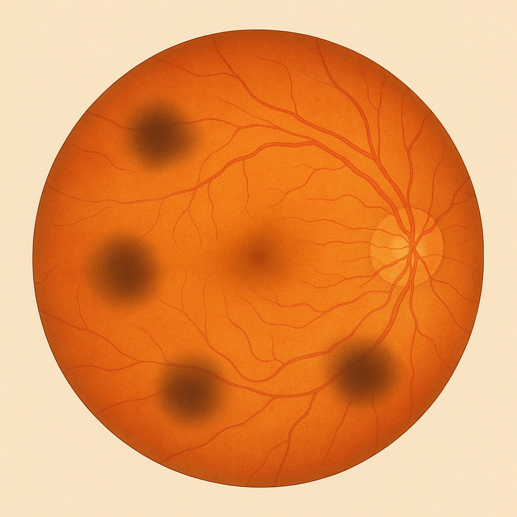
Floaters are small condensations within the vitreous, the transparent gel that fills the inside of the eye. These small opacities cast a shadow on the retina and are perceived as:
- black or gray dots;
- strands or rings;
- small cobwebs that move with the gaze.
They are especially visible against a light background (white wall, sky, computer screen). They do not perfectly follow eye movements but seem to "float" with a slight delay, hence their name floaters.
Why do they appear?
With age, the vitreous changes: it becomes less homogeneous, liquefies, and fibers clump together into small condensations. This normal process explains the majority of floaters in adults.
After age 50, it is common for the membrane surrounding the vitreous to gradually detach from the retina: this is posterior vitreous detachment. This phenomenon is often accompanied by the sudden appearance of new floaters and sometimes flashes of light.
More rarely, floaters can be related to:
- a small vitreous hemorrhage;
- intraocular inflammation;
- a complication of high myopia or trauma.
Floaters: when should you be concerned?
In most cases, isolated floaters that have been present for a long time and are few in number are not dangerous. However, certain situations should raise concern:
- sudden appearance of a shower of floaters in one eye;
- association with flashes of light in the peripheral visual field;
- sensation of a dark veil or a curtain that progresses;
- recent decrease in vision in the affected eye.
These signs may indicate a retinal tear or an early retinal detachment. In this context, floaters are no longer a simple nuisance but a warning symptom.
What the ophthalmologist looks for in an emergency
When a patient presents with recently appearing floaters, the goal is not only to "examine the vitreous" but above all to not miss a retinal lesion. The examination aims to identify:
- a recent posterior vitreous detachment (often the cause of symptoms);
- a peripheral tear (especially if flashes + shower of floaters);
- an early retinal detachment;
- indirect signs such as pigment cells in the vitreous or hemorrhage;
- another rarer cause (inflammation, bleeding, macular traction).
The key point: it is not the presence of a floater that is "serious," but what it may indicate when it is sudden and abundant.
How is the ophthalmological assessment performed?
When floaters are recent or unusual, a comprehensive examination is necessary. The ophthalmologist measures visual acuity, then examines the fundus after pupil dilation. The examination looks for:
- a recent posterior vitreous detachment;
- peripheral retinal tears;
- an early retinal detachment;
- signs of inflammation or bleeding in the vitreous.
In cases of poor media transparency or doubt, an ocular ultrasound may complement the assessment. Macular OCT is useful if traction at the macula, a macular hole, or an associated condition is suspected.
Progression: what happens to floaters over time?
In the vast majority of cases, floaters become less bothersome over time. Two mechanisms explain this:
- the opacities may shift away from the main visual axis;
- the brain adapts and gradually "filters" them out (neuroadaptation).
In some patients, particularly when the opacities are dense or centrally located, the discomfort may persist. In such cases, a personalized discussion is necessary.
What treatments are available for floaters?
When floaters are related to simple aging of the vitreous, there is no medical treatment to make them disappear. Over time, the brain generally learns to ignore them and the discomfort decreases.
Certain techniques (laser vitreolysis or vitrectomy) have been proposed for highly debilitating cases, but they are considered only in exceptional circumstances due to the associated risks. However, if a retinal tear is detected, emergency laser treatment is performed to prevent retinal detachment.
When can treatment be considered?
A specific procedure (laser or surgery) is almost never recommended for ordinary floaters. It may be considered in rare situations when:
- the discomfort is significant and persistent despite several months;
- the opacities are central and very dense;
- the patient is particularly affected in daily life (reading, driving, screen use);
- the examination confirms the absence of retinal pathology requiring priority treatment.
The decision is always made on a case-by-case basis, weighing the expected benefit against the risks.
Daily advice
For most patients, floaters are more of a nuisance than a danger. Some practical tips:
- be reassured: old and stable opacities are generally benign;
- avoid constantly focusing on them, as this increases the discomfort;
- consult promptly if symptoms change suddenly;
- keep the follow-up appointments recommended by your ophthalmologist.
FAQ: floaters
Are floaters always benign?
Most of the time, yes, especially if they are long-standing, few in number, and stable. However, a sudden onset, a large shower, or a rapid increase in floaters should prompt investigation for a retinal tear or even a retinal detachment. The concern is not the symptom itself but what it may sometimes reveal: vitreous traction on the retina during a posterior vitreous detachment.
Why are floaters more visible against a light background?
Because floaters (vitreous opacities) cast a small shadow on the retina. The contrast becomes more noticeable against a white wall, the sky, or a bright screen. Conversely, against a dark or highly textured background, they are often much less noticeable.
Are flashes of light a sign of severity?
They can be. Flashes of light (photopsias) may indicate traction on the retina, particularly during a posterior vitreous detachment. If the flashes are associated with a shower of floaters, black dots "raining down," or a decrease in vision, you should consult promptly to rule out a tear.
Should I seek emergency care if I see a "dark curtain"?
Yes. A dark veil, a curtain that progresses, or an area of the visual field that "goes dark" may correspond to an early retinal detachment. This is an ophthalmic emergency because the earlier the detachment is treated, the lower the risk of macular involvement and visual sequelae.
How long do floaters last?
Floaters can persist for a long time, but the discomfort often decreases within a few weeks to a few months thanks to two phenomena: neuroadaptation (the brain learns to ignore them) and the displacement of opacities away from the visual axis. They may sometimes remain visible under certain conditions (bright light, clear sky), even when they become barely bothersome in daily life.
Can floaters disappear completely?
Sometimes they become virtually invisible in daily life, but they do not always dissolve completely. The primary goal is above all to ensure there is no associated retinal lesion, particularly when symptoms are recent. Once the retina has been checked and if symptoms stabilize, the outcome is most often reassuring.
Are there any effective eye drops or vitamins?
No, there is no recognized medical treatment (eye drops, vitamins) that can eliminate vitreous floaters. The priority is a fundus examination (and sometimes monitoring) to rule out a retinal tear, then allowing time for visual adaptation when the situation is straightforward.
When should a follow-up examination be scheduled?
It depends on the context. After a recent onset, a follow-up may be recommended to ensure no tear develops subsequently, especially in at-risk patients (high myopia, trauma, history of retinal detachment in the other eye). In the meantime, if symptoms worsen (new shower of floaters, more frequent flashes, dark veil, decrease in vision), you should seek care without delay.
When should you consult Julien Gozlan, M.D.?
You should seek a specialist opinion if you suddenly see numerous floaters, if flashes of light accompany them, or if you have the sensation of a veil in your visual field. Individuals with high myopia, those who have previously undergone retinal surgery, or those with a history of retinal detachment should be particularly vigilant.
Julien Gozlan, M.D., ophthalmologist in Paris 16, performs a comprehensive assessment (fundus examination, sometimes ultrasound or OCT) to distinguish benign floaters from forms requiring treatment or close monitoring.
📍 Consultation at Paris – Auteuil Ophthalmology Practice
Julien Gozlan, M.D. welcomes you at Paris – Auteuil Ophthalmology Practice for the diagnosis and follow-up of floaters, posterior vitreous detachment, and associated retinal conditions.
Book an AppointmentFurther reading
- Posterior vitreous detachment: symptoms, risks, and monitoring.
- Retinal detachment: warning signs and management.
- Macular OCT: the key examination for analyzing the retina and the vitreous.