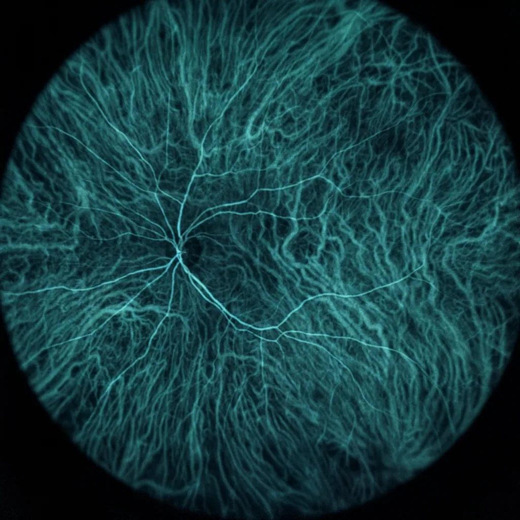

Indocyanine green angiography (ICG) is an imaging examination that highlights the choroidal circulation, located beneath the retina. Complementary to fluorescein angiography and macular OCT, indocyanine green angiography aids in diagnosing choroidal disorders and guides their treatment.
What is indocyanine green angiography?
The examination involves intravenously injecting a dye (indocyanine green or "ICG") that emits fluorescence in the near-infrared spectrum. A specialized camera then records serial images of the choroid and deep vessels. Compared to fluorescein, indocyanine green penetrates pigments more effectively and allows visualization of choroidal abnormalities that may otherwise be invisible.
When should indocyanine green angiography be performed?
This examination is requested when a choroidal disorder is suspected or when other tests are insufficient to explain the symptoms. Common indications include:
- Wet AMD with suspected polypoidal choroidal vasculopathy, occult neovascularization, or chorioretinal anastomosis.
- Central serous chorioretinopathy (CSC) and pachychoroid spectrum disorders.
- Certain types of uveitis and choroidal inflammations.
- Certain choroidal tumors.
- Macular edema of undetermined origin despite OCT and fluorescein angiography.
In these settings, indocyanine green angiography (ICG) maps deep leakage, polyps, and areas of choroidal hyperpermeability, in order to guide management generally toward intravitreal injection therapy, targeted laser treatment, or simple monitoring.
How indocyanine green angiography is performed
Pre-examination questions and preparation
The examination is carried out at the practice. After a few questions (allergies, pregnancy, current medications), the pupils may be dilated using mydriatic eye drops.
Injection of the dye
The indocyanine green dye is injected into a vein in the arm, in the same way as a blood test.
Image acquisition
The camera acquires rapid images at the beginning (early phase), followed by more spaced-out images (intermediate and late phases). Total duration: approximately 25 minutes. A bright light is used; vision may be slightly blurred if dilation has been performed.
End of examination
Indocyanine green angiography is painless: only the venipuncture may cause slight discomfort. Unlike fluorescein angiography, there is no yellow discoloration of the skin or urine. Normal activities can be resumed immediately after the examination.
What indocyanine green angiography reveals
Thanks to this examination, choroidal polyps in polypoidal forms of AMD, choroidal hyperpermeability in CSC, deep neovascularization, and certain inflammatory or tumoral lesions can be visualized. The examination complements fluorescein angiography (which primarily explores the retinal circulation) and OCT to determine the cause of vision loss.
Risks and contraindications
Adverse effects of the examination are rare: transient nausea, vasovagal episode, or reaction at the injection site. The dye contains an iodine derivative: severe allergies are a relative contraindication. The examination is not recommended during pregnancy, except when necessary. The medical team is equipped to manage any unusual reaction.
Interpretation and management
Dr Julien Gozlan correlates indocyanine green angiography images with those from fluorescein angiography and OCT to establish a personalized plan: intravitreal injections, selective laser treatment, or simple monitoring. The goal is to preserve vision and prevent complications.
Frequently asked questions
Does indocyanine green angiography replace fluorescein angiography?
No. Indocyanine green angiography primarily explores the choroid, while fluorescein angiography maps the retinal vessels. The two examinations are complementary.
Can I drive after the examination?
If dilation has been performed, vision will be blurred for a few hours. It is advisable not to drive immediately after indocyanine green angiography.
FAQ: frequently asked questions about indocyanine green angiography (ICG)
Do I need to fast before indocyanine green angiography?
Generally, strict fasting is not required before indocyanine green angiography (ICG), and it is actually preferable to have had a light meal to reduce the risk of vasovagal episodes. However, it is advisable to avoid a very heavy meal just before the examination. If you have a particular medical history (severe allergies, cardiac or renal conditions, pregnancy), specific instructions may be tailored by your ophthalmologist or general practitioner before scheduling the examination.
Is indocyanine green angiography painful or tiring?
The examination is painless from an ocular standpoint: there is no contact with the surface of the eye. The only sensation may come from the venipuncture in the arm, similar to a blood draw. The camera light may seem bright, especially if the pupils are dilated, and the session lasts slightly longer than a simple photograph (approximately 20 to 30 minutes). The vast majority of patients tolerate the examination very well, as it requires no physical effort and is performed in a seated position or slightly reclined in front of the device.
What are the practical differences between ICG angiography and fluorescein angiography?
Both examinations use a dye injected into a vein and a specialized camera, but they do not analyze exactly the same structures. Fluorescein primarily explores the superficial retinal vessels, whereas indocyanine green (ICG) penetrates pigments more effectively and highlights the deep choroidal circulation. In practical terms, ICG causes less discoloration of the skin or urine, and images are captured over a slightly longer period. The choice between the two, or their combination, depends on the type of lesion suspected (polypoidal vasculopathy, central serous chorioretinopathy, particular forms of AMD, etc.) and the specific question the physician wishes to answer.
Is indocyanine green angiography risky in cases of iodine allergy or allergy to certain contrast agents?
Indocyanine green contains an iodine derivative, which requires particular caution in cases of known iodine allergy or allergy to certain agents injected in radiology. This does not necessarily mean the examination is impossible, but the risk of a reaction must be discussed beforehand, sometimes with specific preparation or the choice of an alternative examination. It is therefore essential to inform your ophthalmologist of any drug allergy, history of shock, or severe adverse reaction after an injection, so that the benefit-risk ratio can be assessed and the procedure can be carried out safely if it remains necessary for your care.
Can I drive or work normally after indocyanine green angiography?
If the pupils have been dilated, vision may be blurred and sensitive to glare for a few hours. In that case, it is advisable not to drive immediately after the examination and to avoid visually demanding tasks (prolonged reading, detailed screen work). Normal physical activities can generally be resumed quickly. In practice, many patients arrange to come with a companion or use public transportation, then resume their professional activities the next day or later in the day once vision becomes comfortable again.
How often should indocyanine green angiography be repeated?
Indocyanine green angiography is not an examination that needs to be repeated systematically at fixed intervals. It is primarily performed at the time of initial diagnosis or when a change in disease course or treatment is suspected (change in OCT appearance, new visual decline, suspicion of polyp recurrence or lesion extension). For routine follow-up, OCT and, if needed, OCT angiography are often sufficient. The physician therefore recommends repeating ICG only when the expected information could concretely alter the therapeutic strategy (injections, targeted laser, intensified monitoring, etc.).
Is indocyanine green angiography compatible with pregnancy or breastfeeding?
As a general rule, indocyanine green angiography is avoided during pregnancy, except in exceptional situations where the visual stakes are significant and no other examination can provide the necessary information. Breastfeeding is not an absolute contraindication, but additional precautions may be discussed (for example, pumping and discarding breast milk for a short period after the examination). In all cases, it is essential to inform your ophthalmologist if you are pregnant, planning a pregnancy, or breastfeeding, so that the imaging strategy can be best adapted to your situation.
📍 Indocyanine green angiography at Paris – Auteuil Ophthalmology Practice
Dr Julien Gozlan performs indocyanine green angiography in addition to retinal imaging (OCT, fluorescein angiography) for accurate diagnosis and tailored treatment.
Book an AppointmentFurther reading
- Fluorescein angiography: mapping retinal vascular disorders.
- Age-related macular degeneration (AMD): the role of imaging in follow-up.
- Intravitreal injections: targeted treatments guided by imaging.