Retinal Surgery
13 articles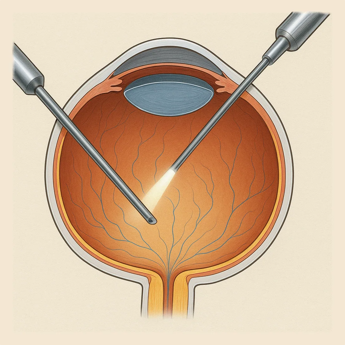
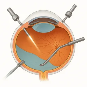
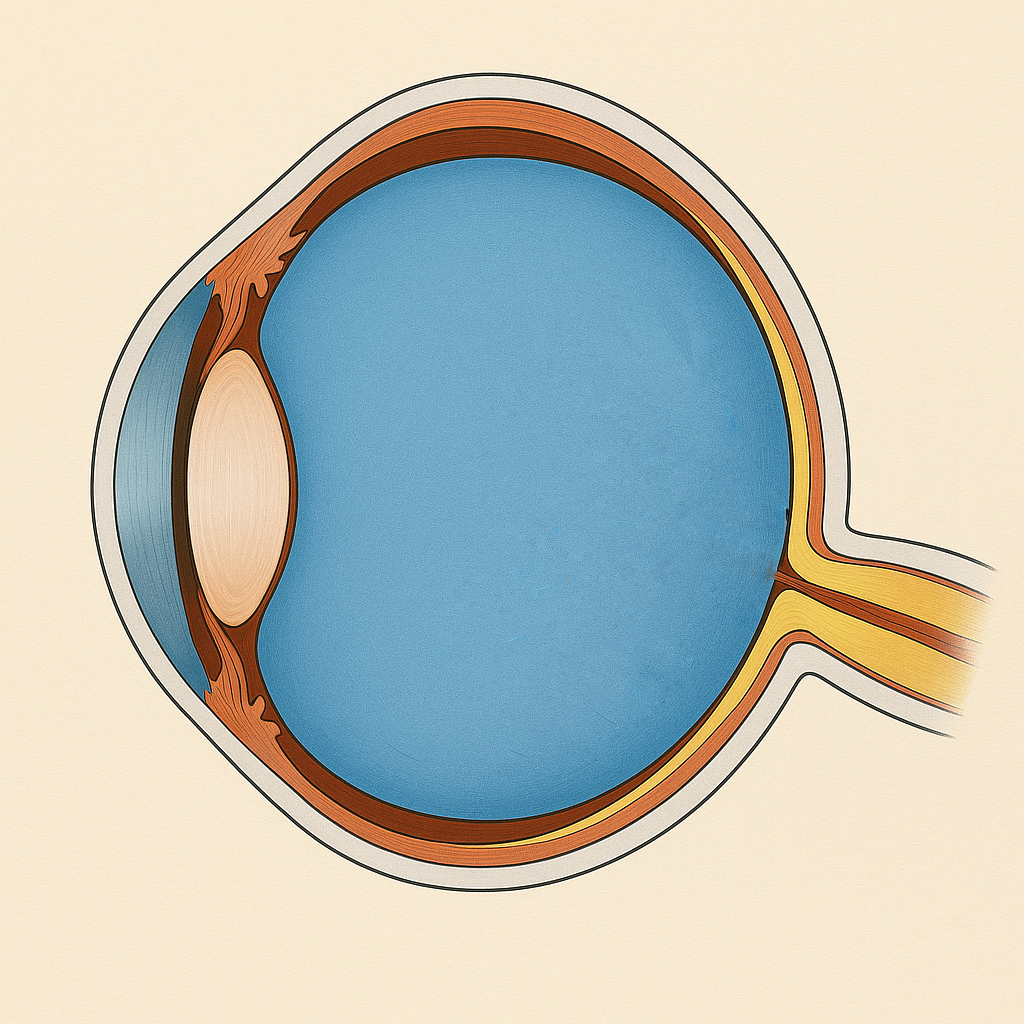
Vitrectomy in vitreoretinal surgery
Vitrectomy is an eye surgery that involves removing the transparent gel called the vitreous, located in the center of the eye. It is performed to treat certain retinal diseases that may threaten vision. Julien Gozlan, M.D., ophthalmic surgeon in Paris 16, explains the principle of this surgery, its main indications, and how the procedure is carried out.
Read more
Macular Epiretinal Membrane
A macular epiretinal membrane is a thin layer of tissue that forms on the surface of the macula, the central area of the retina responsible for detailed vision. It can cause image distortion (metamorphopsia), progressive vision loss, and difficulty reading. It most commonly appears after the age of 50, related to natural changes in the vitreous. Julien Gozlan, M.D., ophthalmic surgeon in Paris 16, explains the causes, symptoms, the role of OCT, and the surgical treatment.
Read more
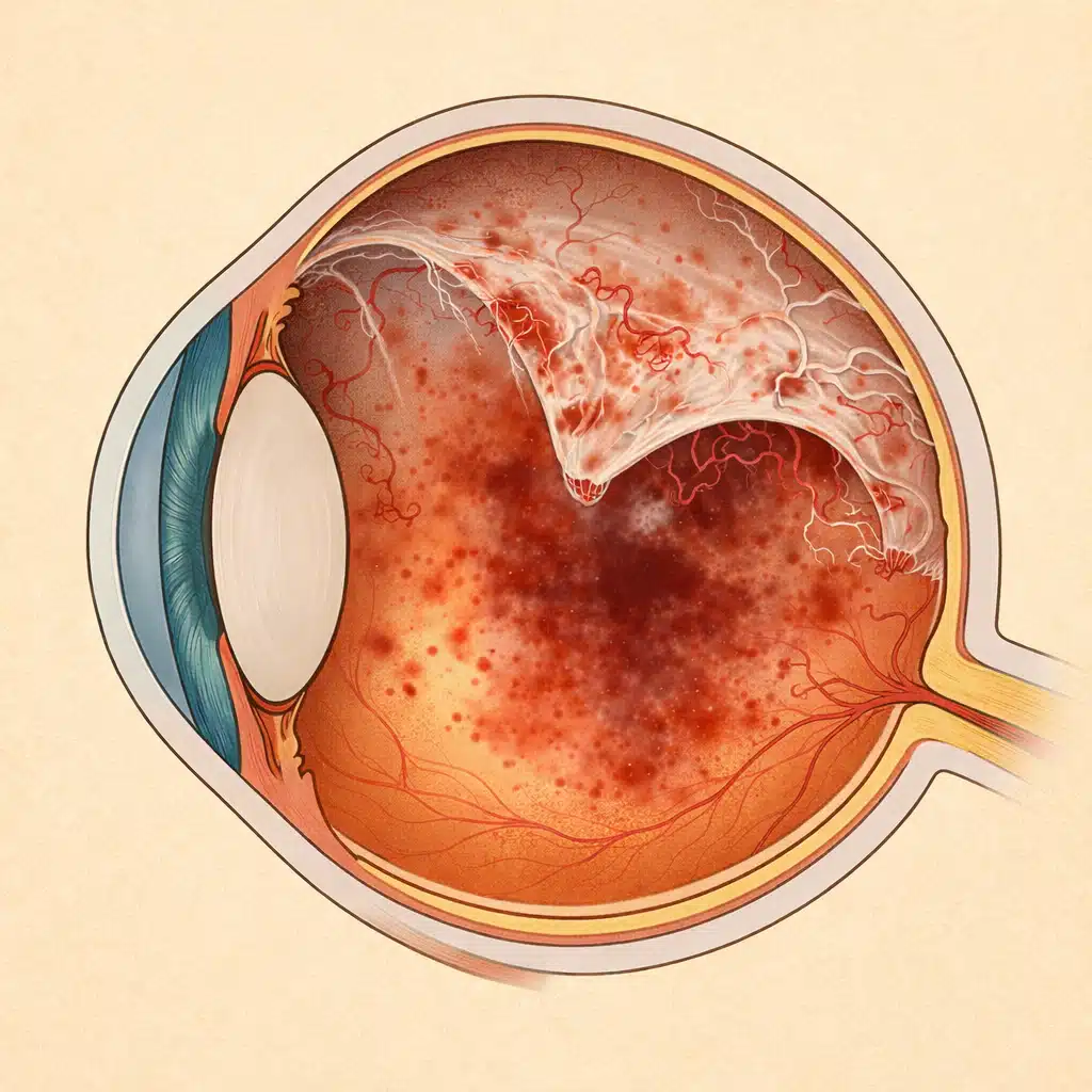
Retinal Detachment
Retinal detachment is an ophthalmological emergency: the retina lifts away and no longer receives light properly, putting vision at risk. Prompt treatment significantly improves the chances of recovery. Julien Gozlan, M.D. explains the warning symptoms, diagnosis, and main surgical options to preserve your sight.
Read more
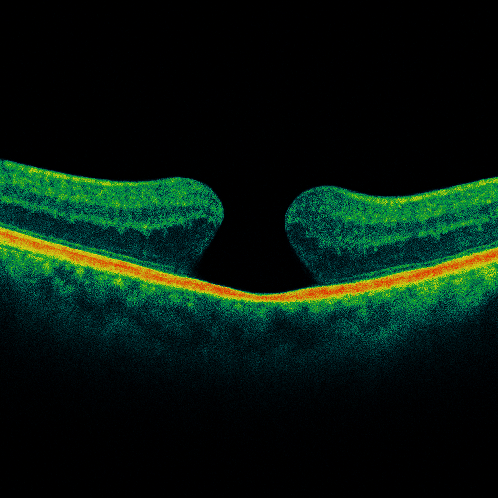
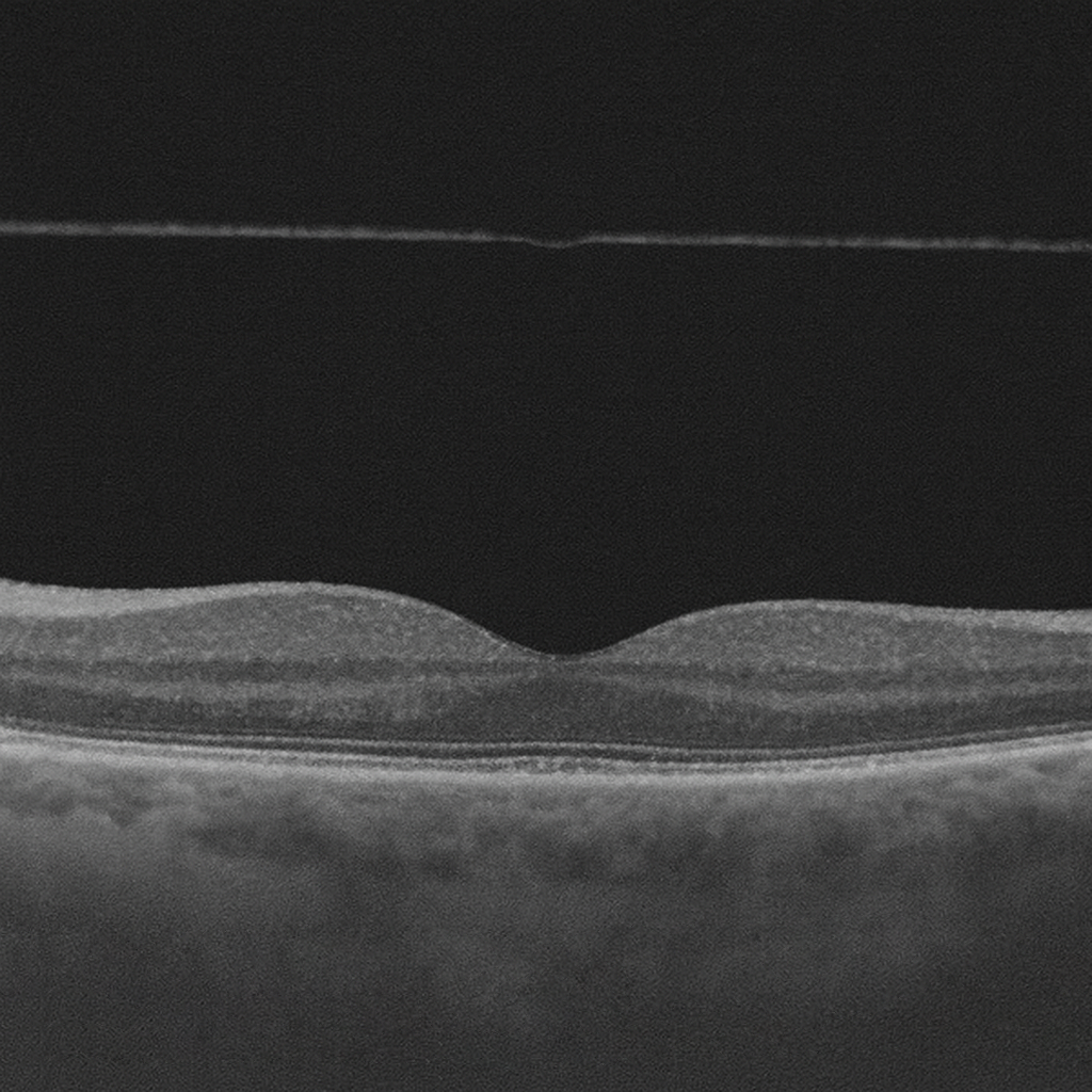
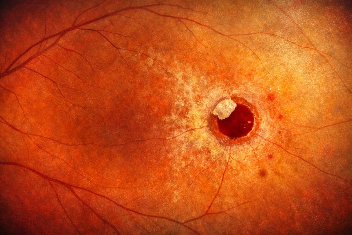
Macular Hole
A macular hole is a small opening in the center of the retina, within the macula. This condition often causes decreased central vision, a spot in the middle of the visual field, and distorted lines. Julien Gozlan, M.D., ophthalmic surgeon in Paris 16, explains the OCT diagnosis, treatment options, and prognosis.
Read more
Retinal Detachment Surgery
Retinal detachment surgery is an ophthalmic emergency: without prompt treatment, vision can be permanently lost. Retinal detachment surgery relies primarily on two techniques: cryopexy with scleral buckling and vitrectomy. Julien Gozlan, M.D., ophthalmic surgeon in Paris 16, explains the differences between these procedures, their indications, and postoperative recovery.
Read more
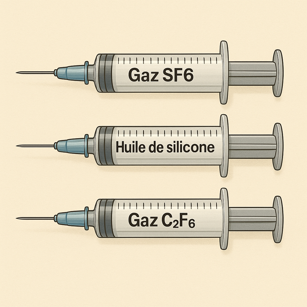
Vitrectomy and Tamponade
During vitreoretinal surgery, the surgeon may fill the eye with an intraocular tamponade on a temporary or permanent basis. This temporary «filling» of the eye is performed using air, gas, or silicone oil, depending on the situation. Julien Gozlan, M.D., ophthalmic surgeon in Paris 16, details the main types and their indications.
Read more
Posterior Vitreous Detachment
Posterior vitreous detachment is a very common condition after the age of 50. The transparent gel that fills the eye, the vitreous, gradually separates from the retina. In most cases, the phenomenon is benign, but it can sometimes be accompanied by a retinal tear.
Read more
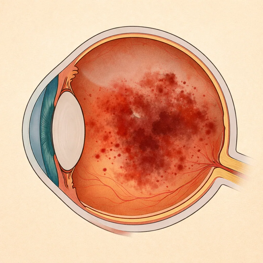
Vitreous Hemorrhage
Vitreous hemorrhage refers to the presence of blood within the vitreous, the transparent gel that fills the interior of the eye. It often causes sudden vision loss, the appearance of a dark veil, or a «shower of soot» effect. Some forms are straightforward and resolve spontaneously, while others may conceal a retinal detachment or severe vascular disease. Julien Gozlan, M.D., ophthalmic surgeon in Paris 16, explains the causes, symptoms, diagnostic examinations (OCT, ultrasound) and treatments for vitreous hemorrhage.
Read more
Vitrectomy and Diabetic Retinopathy
Diabetic retinopathy is a common and potentially severe complication of diabetes. When recurrent vitreous hemorrhages occur or vascular proliferation becomes significant, a vitrectomy may be necessary to save or stabilize vision. Julien Gozlan, M.D., ophthalmic surgeon in Paris 16, explains the role of vitrectomy in diabetic retinopathy, its indications, how the procedure is performed, and the visual prognosis.
Read more
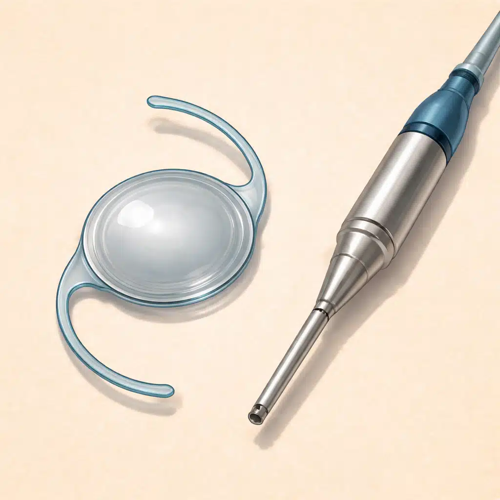
Combined cataract and vitrectomy surgery
Combined surgery allows both a cataract and a retinal or vitreous condition (epiretinal membrane, macular hole, diabetic retinopathy, retinal detachment, etc.) to be treated in a single operation. It avoids two separate procedures and shortens recovery time, but requires precise planning and choices tailored to each case. Julien Gozlan, M.D., ophthalmic surgeon in Paris 16, explains the indications, procedure, and prognosis of this combined surgery.
Read more
Retinal tear
A retinal tear is a break in the retinal tissue, often caused by traction from the vitreous (the gel that fills the eye). It can remain silent for a long time or present with floaters, flashes of light, or a curtain effect in the visual field. The main risk is progression to a retinal detachment, which constitutes a surgical emergency. Julien Gozlan, M.D., ophthalmologist in Paris 16, explains the warning symptoms, diagnosis, laser treatments, and the follow-up that should be implemented.
Read more
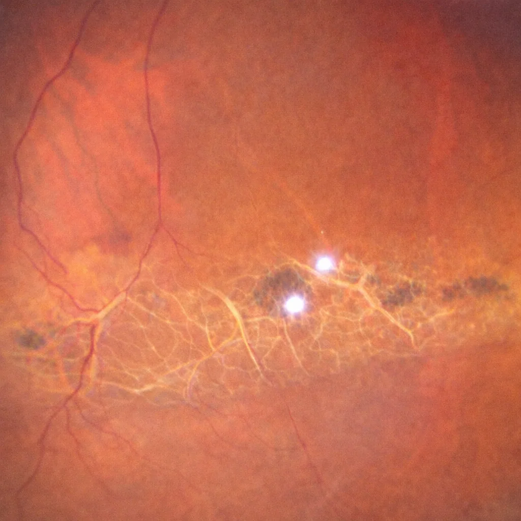
Lattice Degeneration
Lattice degeneration, also known as lattice retinal degeneration, refers to thin, weakened areas of the peripheral retina, often discovered during a routine fundus examination. It is generally asymptomatic but can increase the risk of retinal tears and subsequent retinal detachment in certain specific situations. Julien Gozlan, M.D., ophthalmologist in Paris 16, explains what lattice degeneration is, when it should simply be monitored, and in which cases prophylactic laser retinopexy is truly indicated.
Read more
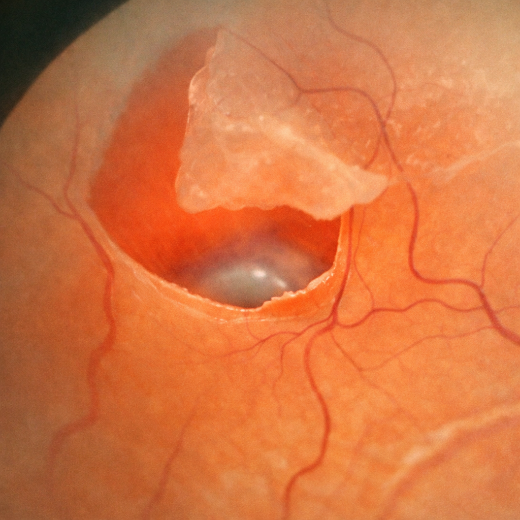
Operculated retinal hole
An operculated hole is a lesion of the peripheral retina caused by a posterior vitreous detachment (PVD). During this physiological process, the vitreous exerts traction on the retina and tears away a small fragment of retinal tissue, called an operculum, which remains suspended above the hole. This lesion is managed either through simple monitoring or preventive laser retinopexy performed in the office; surgery is only indicated in cases of associated retinal detachment. Julien Gozlan, M.D., ophthalmologist in Paris 16, explains in this article the mechanisms of formation, symptoms, diagnosis, and management of this peripheral retinal lesion.
Read more