
Retinal detachment is an ophthalmological emergency: the retina lifts away and no longer receives light properly, putting vision at risk. Prompt management significantly improves the chances of recovery. Julien Gozlan, M.D. explains the warning symptoms, the diagnosis, and the main surgical options to preserve your sight.
What is retinal detachment?
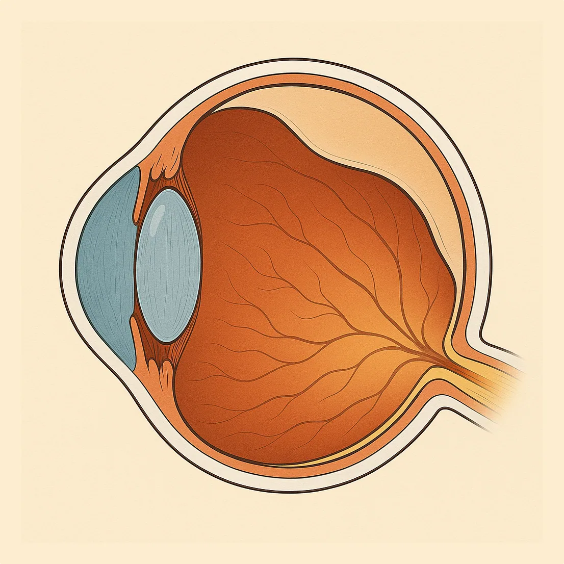
Retinal detachment occurs when the neurosensory retina separates from the layer that nourishes it (retinal pigment epithelium). Deprived of oxygen, the retina functions less effectively: vision loss can become irreversible if intervention is delayed too long.
Most often, it all begins with a retinal tear: fluid passes beneath the retina and progressively detaches it, much like wallpaper peeling away from a wall. The options for retinal detachment surgery depend on the extent, the location of the tears, and the condition of the vitreous.
Retinal detachment: warning symptoms
The key sign is often the sudden onset of visual symptoms in one eye. The most typical warning signs are:
- flashes of light (photopsias), especially in the dark;
- floaters or sudden, numerous black spots;
- dark curtain (veil) progressing across the visual field;
- vision loss or distortion if the macula is threatened.
If you experience these signs, seek emergency care: a retinal detachment detected early is easier to treat and often yields a better outcome.
What are the causes and risk factors of retinal detachment?
In the majority of cases, it is a rhegmatogenous retinal detachment: a tear allows fluid to pass beneath the retina. The main risk factors are:
- myopia, especially high myopia;
- age (posterior vitreous detachment);
- history of detachment in the fellow eye;
- ocular trauma;
- certain surgeries (notably cataract surgery in certain patient profiles).
In highly myopic patients, the retina is thinner and more stretched, which increases the risk of tears. Certain macular conditions associated with myopia (such as myopic tractional maculopathy) can also weaken the central area and complicate follow-up.
How is the diagnosis confirmed?
The diagnosis is based on a dilated fundus examination. It allows the tear or tears to be located and the extent of the detachment to be assessed.
If the fundus is difficult to visualize (vitreous hemorrhage, dense cataract, miosis), an ocular ultrasound can confirm the presence of a retinal detachment.
A macular OCT is sometimes performed if the macula is threatened or to clarify the macular status, which helps estimate the prognosis.
OCT signs and key findings
When OCT is performed, it provides useful information about any central involvement from the retinal detachment:
- macula status: macula "on" (still attached) or macula "off" (already detached);
- presence of subretinal fluid at the posterior pole;
- possible associated vitreomacular traction;
- signs of macular fragility (high myopia, membranes, retinoschisis).
In practice, the essential question is: is the macula involved? This is a major determinant of visual prognosis.
Treatment: what surgical options are available?
Treatment always aims to reattach the retina and to seal the tear (laser or cryotherapy). The choice depends on the type of detachment, the tears, the patient's age, the degree of myopia, and the condition of the vitreous. In some cases, several techniques are combined.
Vitrectomy (surgery from the inside)
Vitrectomy involves removing the vitreous, releasing traction, and then treating the tears with laser (or cryotherapy). The eye is then filled with a gas or silicone oil to keep the retina reattached during healing. It is currently one of the most widely used techniques in retinal surgery.
Scleral buckle (surgery from the outside)
Scleral buckling involves placing a silicone band around the eye (or a local implant) to reduce traction on the tear. This technique is often preferred in young (phakic) patients with an accessible peripheral tear, depending on the clinical context.
Intraocular gas and positioning
When a gas tamponade is used, specific positioning may be required for several days (for example, face down) so that the bubble presses against the treated area. Instructions are tailored to the location of the tears and the type of gas used.
Postoperative course: what you need to know
After surgery, eye drops are prescribed (anti-inflammatory and antibiotic) and frequent follow-up visits are scheduled. When gas is present, vision is blurry at first and then gradually improves.
Air travel and nitrous oxide anesthesia are contraindicated as long as gas remains in the eye, because the bubble can expand and cause a dangerous rise in intraocular pressure.
Prognosis: what determines recovery?
The prognosis depends mainly on the timeliness of treatment and the macular status. A retinal detachment operated on promptly before the macula is affected generally produces the best results. If the macula has been detached, the retina can be reattached but visual recovery is often slower and sometimes incomplete.
Recurrences are possible, particularly in cases of proliferative vitreoretinopathy or new tears. Regular follow-up allows early detection of any disease progression. For reliable patient information, you may consult the Retina France article on retinal detachment.
FAQ: frequently asked questions about retinal detachment
Is it always an emergency?
Yes. Even if vision remains adequate initially, the risk of the detachment extending is real. A prompt ophthalmological assessment is essential to confirm the diagnosis and plan appropriate management.
Why do you see flashes of light?
Flashes of light (photopsias) often indicate mechanical traction on the retina, typical of a posterior vitreous detachment that may be accompanied by a retinal tear. They are a warning sign that should prompt an urgent consultation.
Do floaters necessarily mean a detachment?
No. Floaters can be benign, but the sudden appearance of a large shower of floaters, especially when associated with flashes of light or a curtain effect, should prompt an emergency visit to rule out a tear or retinal detachment.
Can surgery be avoided?
When the retina is already detached, surgery is the standard treatment. However, certain isolated tears, discovered early before a detachment has developed, can sometimes be treated with laser (or cryotherapy) without the need for vitrectomy or more extensive surgery.
Do I need to maintain a specific position after the operation?
Sometimes yes, especially when intraocular gas has been injected. The head position (for example, face down into a pillow or on one side) is chosen so that the gas bubble presses precisely on the treated area. Instructions vary depending on the location of the tears and the type of surgery performed.
When can I resume driving and work?
This depends on the type of procedure, whether or not gas is still present in the eye, the visual recovery, and the type of work involved. Driving is only permitted once vision and visual field are deemed sufficient. Your surgeon will provide personalized recommendations during follow-up appointments.
Why is air travel prohibited when intraocular gas is present?
At altitude, cabin pressure decreases and the gas bubble inside the eye expands. This can cause a sudden and severe rise in intraocular pressure, which is potentially dangerous for the eye. Until the gas has been completely resorbed, air travel (and certain general anesthetics using nitrous oxide) is strictly contraindicated.
Is there a risk for the fellow eye?
Yes, the risk is somewhat higher in the fellow eye, especially in cases of high myopia, family history, or fragile peripheral retinal lesions. Regular monitoring of the fellow eye, including a dilated fundus examination, is often recommended to detect any tear or retinal abnormality early.
📍 Consultation at the Paris – Auteuil Ophthalmology Practice
Julien Gozlan, M.D. manages retinal detachment: diagnosis, emergency surgical treatment (vitrectomy, scleral buckle), and personalized follow-up to optimize long-term visual outcomes.
Book an AppointmentFurther reading
- Retinal detachment surgery: cryo-buckle or vitrectomy, how to choose.
- Vitrectomy: a major surgical technique in vitreoretinal surgery.
- Macular OCT: useful for evaluating the macula before and after surgery.