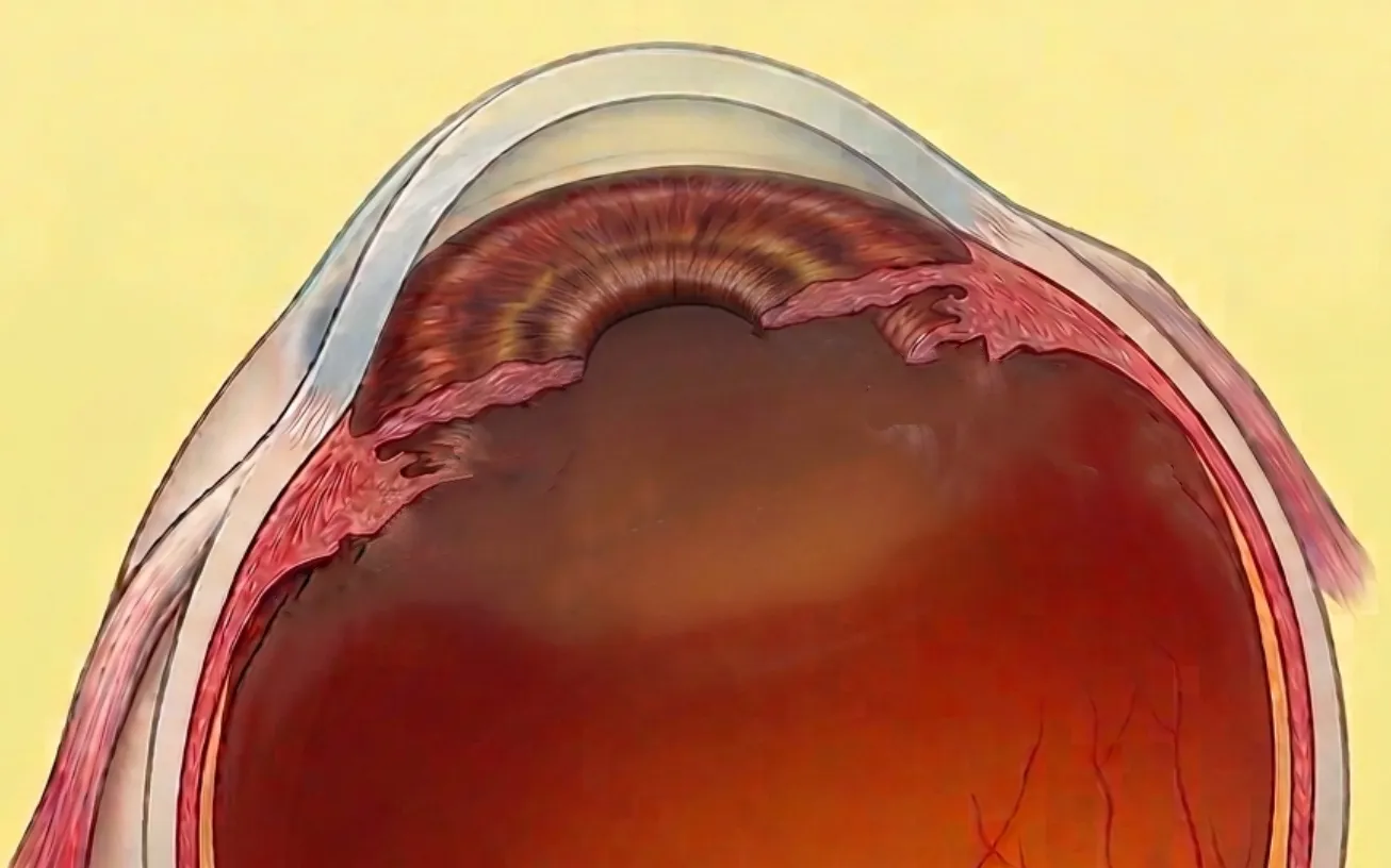
Secondary IOL implantation means placing an intraocular lens in an eye that has lost the natural structure meant to hold it: the capsular bag. This secondary IOL implantation becomes necessary after a dislocated lens, a capsular rupture that led the surgeon to leave the eye aphakic, or long-standing aphakia. Several techniques coexist today — iris-claw fixation, iris suture, scleral suture, Yamane intrascleral fixation, Carlevale lens — and they differ markedly in difficulty, in track record and in complication profile. Dr Julien Gozlan, ophthalmic surgeon specialising in anterior segment and vitreoretinal surgery in Paris 16, sets out what genuinely separates these options.
What is secondary IOL implantation?
In routine cataract surgery the intraocular lens is placed inside the capsular bag, a transparent envelope suspended by several hundred microscopic fibres, the zonule of Zinn. When that support is absent, torn or too weak, the lens can no longer sit there: this is secondary IOL implantation, an implantation carried out at a later stage and using a different anchoring point.
A secondary IOL implantation therefore differs from an ordinary one in a single respect, but a decisive one: something else must hold the lens. Three structures can provide that hold — the iris, the sclera, and more rarely the iridocorneal angle. That choice of anchor is what defines the technique of secondary IOL implantation.
When is secondary IOL implantation indicated?
The situations that lead to secondary IOL implantation are well established:
- Late lens dislocation: the capsular bag, weakened by zonular fragility, pseudoexfoliation or high myopia, eventually shifts along with the lens it carries;
- Intraoperative capsular rupture: when the posterior capsule gives way during surgery and no reliable support remains, the surgeon often prefers to defer implantation;
- Aphakia: an eye left without a lens after trauma or older surgery;
- Subluxated crystalline lens: Marfan syndrome, homocystinuria, blunt trauma;
- Poorly tolerated lens that needs replacing.
The decision never rests on anatomy alone. A secondary IOL implantation is weighed first on the actual handicap: reduced vision, monocular double vision, halos, or an associated complication such as raised pressure or inflammation.
The work-up before secondary IOL implantation
No secondary IOL implantation is planned without a full work-up, which governs the choice of technique as much as the visual outcome:
- ocular biometry, essential to recalculate lens power in its new position;
- endothelial cell count, which weighs heavily: a fragile cornea rules out anything sitting too far forward;
- a macular OCT scan, looking for pre-existing oedema;
- careful assessment of the iris, which alone decides whether a clip-based secondary IOL implantation is feasible — its tone, its integrity over 360°, the roundness of the pupil;
- examination of the peripheral retina, since the risk of retinal detachment rises as soon as the vitreous is handled.
Iris-claw lens: the benchmark in secondary IOL implantation
The iris-claw lens was born in 1977-1978 in the hands of the Dutch surgeon Jan Worst. It rests on a simple idea, and it remains the most widely used approach in secondary IOL implantation: two lateral claws enclavate a fold of iris stroma at 3 and 9 o'clock. Since the iris carries few vessels at these points, it tolerates the grip remarkably well. Close to 300,000 aphakic eyes have been implanted this way worldwide, which makes it by far the secondary IOL implantation option with the longest clinical track record.
Fixation is now almost always retropupillary: the lens is slipped behind the iris and enclavated from underneath. This variant moves the optic away from the cornea, protects the endothelium and leaves the iridocorneal angle untouched. It has become the standard for clip-based secondary IOL implantation.
The published figures for this secondary IOL implantation are robust. In a series of 103 eyes followed for a mean of 38 months, corrected acuity gained 0.69 logMAR within the first month and remained stable thereafter. Endothelial cell loss reached 16.6% at one month and then plateaued, with no bullous keratopathy at three years. The complications are familiar and manageable: disenclavation 9.7% (a short revision), pupil ovalisation 10.7%, transient pressure rise 7.8%, cystoid macular oedema 3.9%, and no retinal detachment.
How is secondary IOL implantation with an iris-claw lens performed?
Surgery takes place in theatre, usually under locoregional anaesthesia, as a day case. A clip-based secondary IOL implantation lasts twenty to forty minutes.
1. Preparation and vitrectomy
If vitreous has come forward into the anterior chamber, or if the old lens has fallen back, a pars plana vitrectomy is performed first. This step matters: leaving vitreous around the construct invites retinal traction.
2. Removal of the dislocated lens
The unstable lens is lifted and extracted through a corneal incision, or cut so that it can leave through a smaller opening.
3. Placement behind the iris
The new lens is introduced and positioned behind the iris plane, with the optic centred on the visual axis.
4. Enclavation
An enclavation needle pushes a fold of iris into each claw. This decisive manoeuvre governs the centration and stability of the entire secondary IOL implantation.
5. Checks and closure
The surgeon confirms a round pupil, the absence of incarcerated vitreous, and watertight incisions, suturing them where needed.
Iris-sutured lens: a declining form of secondary IOL implantation
Iris suture fixation, described by McCannel, secures the haptics of a three-piece lens to the iris stroma with 10-0 polypropylene, the optic sitting behind the iris. Technically more accessible than scleral fixation, it served well for many years.
It is far less used today. Suturing hardware to uveal tissue — richly vascularised and mobile with every change in light — invites chronic inflammation, pigment dispersion and the feared uveitis-glaucoma-hyphaema syndrome. Some series report up to 28% macular oedema. This form of secondary IOL implantation is now reserved for perfectly round, well-toned irides.
Scleral-sutured lens: solid, but the sutures age
Here the anchor leaves the iris for the sclera. Haptics carrying eyelets are tied to the scleral wall with a suture passed ab externo — 9-0 or 10-0 polypropylene, or the sturdier 8-0 Gore-Tex. The appeal is obvious: every eye has a sclera, including those whose iris is atrophic or missing.
The weak point is the suture itself. Polypropylene breakage eight to fifteen years after placement is documented, as is knot erosion, a portal for endophthalmitis. A four-eyelet lens is more stable than a two-eyelet one, and Gore-Tex, thicker and less prone to cheese-wiring, has largely replaced fine polypropylene in units that perform this construct regularly. This secondary IOL implantation also takes noticeably longer.
Yamane technique: sutureless intrascleral fixation
Described in 2017, the Yamane technique is the secondary IOL implantation that does away with sutures. Both haptics of a three-piece lens are externalised through the sclera using thin 27- or 30-gauge needles, then their tips are melted with low-temperature cautery into a small mushroom head — the flange — which locks inside a scleral tunnel.
A meta-analysis of 13 studies and 737 eyes places it level with scleral suturing on final acuity, macular oedema and retinal detachment, while cutting roughly 25 minutes of operating time. Its limits lie in execution: the needle tracks are made blind, centration is demanding, residual tilt is hard to correct afterwards, and haptic material is critical — a brittle haptic risks fracture. It also requires lenses used outside their official indication.
Carlevale lens: self-anchoring scleral fixation
The Carlevale lens (FIL SSF) was designed for this purpose alone. A single-piece hydrophilic acrylic lens with a 6.5 mm optic, it carries two T-shaped harpoons that cross the sclera and anchor themselves inside scleral pockets, with neither suture nor glue, and four counter-pressure points that limit tilt.
Published results are encouraging: across 169 eyes, corrected acuity improved from 0.58 to 0.09 logMAR at nine months, with no reported tilt or decentration and a lower retinal detachment rate than other constructs. Against that stand a frequent postoperative pressure rise (16.5%), macular oedema in 7.4%, reports of optic calcification, and scleral pocket thinning that warrants monitoring. This secondary IOL implantation is still young: its track record is measured in years, not decades.
Which technique for secondary IOL implantation?
Meta-analyses comparing iris-claw fixation with scleral fixation give similar visual results: the American Academy of Ophthalmology report (2020) finds no superiority of any technique, and the most recent meta-analysis (2024, 534 eyes), which finds a slightly better final acuity with sutureless scleral fixation, reports a similar complication rate and a markedly longer operating time with the latter. Iris-claw fixation remains the shortest, simplest and quickest procedure to learn.
With visual outcomes similar, what separates one secondary IOL implantation from another is durability over time. On that ground, the classic retropupillary iris-claw lens remains the most proven and the safest technique: forty years of follow-up, hundreds of thousands of eyes operated, a short and reproducible procedure, a construct that can be repositioned if it disenclavates, and a fully mapped complication profile. None of the newer techniques can put forward a comparable track record today.
That does not disqualify them. When the iris is atrophic, torn or absent, no iris grip is possible and scleral fixation — Yamane or Carlevale — becomes the right answer. The choice is made eye by eye, but against a clear benchmark.
FAQ: secondary IOL implantation
Is secondary IOL implantation done under general anaesthesia?
Rarely. The great majority of secondary IOL implantation procedures are performed under locoregional anaesthesia as a day case. The eye is completely numb and still; general anaesthesia is kept for very anxious patients or those unable to lie flat.
How long does vision take to recover?
After a clip-based secondary IOL implantation, useful vision returns within days and settles over six to eight weeks. Scleral fixation often asks for a little more patience. Macular oedema or inflammation can delay that recovery.
Can the lens move again after surgery?
It can, but it is uncommon. Disenclavation of an iris-claw lens occurs in about 10% of cases and is corrected by a short revision. Suture-based fixation can give way much later, sometimes after ten years.
Does the dislocated lens have to be removed?
Usually yes: during a secondary IOL implantation, a single-piece acrylic lens lends itself neither to suturing nor to scleral fixation. Some three-piece lenses in good condition can, however, be repositioned rather than exchanged.
What are the risks of secondary IOL implantation?
The main ones are macular oedema, transient pressure rise, endothelial cell loss, pupil ovalisation and, more rarely, retinal detachment or endophthalmitis. These risks remain low and are monitored at postoperative visits.
When should you see Dr Julien Gozlan about secondary IOL implantation?
Any drop in vision, distortion of images or double vision in one eye in a patient already operated for cataract warrants examination. A slit-lamp examination through a dilated pupil identifies a displaced lens within minutes and clarifies the state of the capsular support and the iris — the two elements that steer the secondary IOL implantation.
Dual expertise in lens surgery and vitreoretinal surgery matters here: it allows the vitreous, the retina and the lens to be dealt with in a single operation, sparing the patient a second procedure.
📍 Consultation at the Paris – Auteuil Ophthalmology Practice
Dr Julien Gozlan sees patients at the Paris – Auteuil Ophthalmology Practice for the assessment of a displaced intraocular lens and the surgical management of secondary IOL implantation.
Book an appointment on DoctolibFurther reading
- Dislocated intraocular lens: why a lens moves and when surgery becomes necessary.
- Intraocular lenses: monofocal, toric, multifocal — understanding the artificial lens.
- Vitrectomy: the retinal step that so often accompanies these procedures.
- Ocular biometry: how lens power is recalculated.
- Combined surgery: treating the lens and the retina in one sitting.