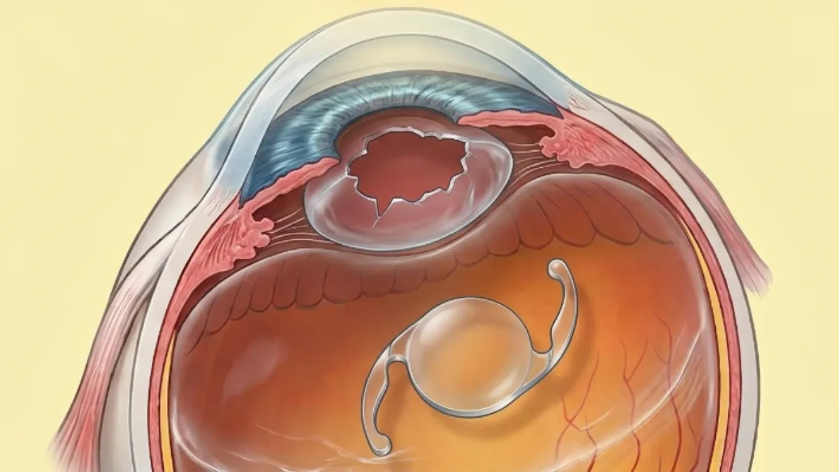

A dislocated intraocular lens after cataract surgery means that the artificial lens has migrated out of its normal position inside the capsular bag. This complication is rare, but it is well documented and entirely treatable: a dislocated intraocular lens may occur a few days, or fifteen years, after the operation. Dr Julien Gozlan, ophthalmic surgeon specialising in anterior segment and vitreoretinal surgery at the Cabinet Ophtalmologique Paris – Auteuil, explains it all: mechanisms, risk factors, symptoms, confirmatory tests, surgical repositioning techniques and visual prognosis.
What is a dislocated intraocular lens after cataract surgery?
Cataract surgery consists of removing the clouded natural lens and replacing it with an intraocular lens, an artificial lens slipped into the capsular bag that is left in place. That bag, extremely thin, is suspended from the eye wall by several hundred microscopic fibres: the zonule of Zinn. We speak of a dislocated intraocular lens when this assembly leaves its position centred on the visual axis.
Surgeons distinguish two situations. Intraocular lens subluxation is a partial displacement: the lens decentres and tilts, but its edge remains visible within the pupil. Complete lens dislocation means the lens has fallen entirely, usually backwards into the vitreous cavity, where it rests on the retina. Between these two extremes lies a whole continuum, and it is precisely the degree of decentration of a dislocated intraocular lens that guides management.
A dislocated intraocular lens must also be distinguished from two far more common and unrelated situations: clouding of the posterior capsule, or secondary cataract, which is treated with a laser in a few minutes, and a lens power error, which stems from an inaccuracy in the preoperative ocular biometry.
Why does a dislocated intraocular lens occur? The main causes
In the vast majority of cases, a dislocated intraocular lens reflects failure of the support holding the lens, not a defect of the intraocular lens itself. The causes break down as follows:
- Zonular weakness: this is the dominant cause of late cases. Pseudoexfoliation syndrome, a progressive deposit of fibrillar material on the structures of the eye, weakens the zonule and multiplies the risk of a dislocated intraocular lens by five to ten.
- Ocular trauma: a direct blow, even several years after surgery, can abruptly rupture the remaining zonular fibres.
- High myopia and long axial length: a large eye is exposed to increased zonular and capsular fragility.
- Previous surgery: a prior vitrectomy, glaucoma surgery or a posterior capsular rupture occurring during cataract surgery markedly increase the risk.
- Capsular contraction: excessive fibrosis of the capsular bag, known as capsular phimosis, can literally pull the lens out of its axis.
- Certain systemic or ocular diseases: Marfan syndrome, homocystinuria, chronic uveitis, retinitis pigmentosa.
Dislocated intraocular lens: which symptoms should raise the alarm?
The signs depend entirely on the extent of decentration. A dislocated intraocular lens of a few tenths of a millimetre may remain silent for a long time, whereas a complete dislocation causes a sudden collapse of vision.
- Blurred vision, gradual or sudden, that glasses no longer correct;
- Double vision in one eye (monocular diplopia), highly suggestive of a dislocated intraocular lens;
- Halos, glare and reflections around light sources, caused by light passing over the edge of the lens;
- The sensation of a moving veil or a dark crescent at the edge of the visual field;
- Vision that changes with head position, an almost specific sign of a mobile lens;
- Suddenly very blurred vision, as if the eye had never been operated on at all, in cases of complete dislocation into the vitreous.
Any of these signs in a patient who has had cataract surgery, however long ago, warrants prompt ophthalmic examination: a dislocated intraocular lens can be complicated by raised eye pressure, inflammation or retinal detachment.
Early or late dislocated intraocular lens: two distinct mechanisms
Timing strongly guides the diagnosis. A so-called early dislocated intraocular lens, occurring within the first three months, almost always reflects a capsular support problem created during surgery: posterior capsule rupture, intraoperative zonular dialysis, or implantation into an incomplete bag. The lens then slides within a capsule that can no longer retain it.
A late dislocated intraocular lens, appearing after several years, corresponds to a very different mechanism: the capsular bag and lens migrate together as a single block, because the zonule has gradually weakened. This "in-the-bag" scenario is overwhelmingly associated with pseudoexfoliation syndrome. The mean time to onset is around eight to ten years after surgery, and the cumulative incidence remains low, of the order of 0.1 to 1% at twenty years.
Which tests confirm a dislocated intraocular lens?
The diagnosis of a dislocated intraocular lens rests above all on slit-lamp examination through a dilated pupil. The surgeon assesses the exact position of the lens, the integrity of the capsular bag, iris mobility (phacodonesis and iridodonesis) and the state of the zonule over 360 degrees.
Depending on the case, this examination is completed by a dilated fundus examination, essential to locate a lens that has fallen into the vitreous and to look for an associated retinal tear, by macular optical coherence tomography (OCT) looking for oedema, by intraocular pressure measurement, and by B-scan ultrasound when the fundus cannot be seen. Finally, biometry is performed if a lens exchange is being considered.
Does a dislocated intraocular lens always need surgery?
No, and this is an essential point. The decision depends on the actual visual impact, not solely on the appearance at examination. A minimal, stable dislocated intraocular lens, without functional disturbance or complication, may simply be monitored, with regular checks on lens position, intraocular pressure and macular status.
Conversely, surgical revision is required in the face of significant visual loss, disabling monocular diplopia, a lens that has fallen into the vitreous, ocular hypertension, chronic inflammation or corneal damage from rubbing. In a patient with only one functioning eye, surgery is indicated more readily and earlier.
Surgery for a dislocated intraocular lens: repositioning techniques
Surgical management of a dislocated intraocular lens is carried out in the operating theatre, most often under locoregional anaesthesia, as a day case. The strategy depends on the position of the lens, the quality of the remaining capsular support and the type of intraocular lens in place.
- Simple intraocular lens repositioning: when the capsular bag is still partly supported, the lens can be recentred and stabilised with a capsular tension ring, if necessary fixated to the sclera.
- Scleral or iris suture: the existing lens is anchored with a non-absorbable suture to the eye wall or to the iris, which avoids having to remove it.
- Sutureless scleral fixation: modern techniques such as the Yamane or glued IOL methods allow the haptics of the lens to be tucked into scleral tunnels, with excellent stability results.
- Lens exchange: the lens is removed and replaced, in particular by an iris-fixated anterior chamber intraocular lens when no posterior support can be used.
- Associated vitrectomy: essential as soon as the lens has fallen into the vitreous, it allows safe retrieval and treatment of any retinal lesion. This is the principle of combined surgery.
Recovery and visual prognosis after a dislocated intraocular lens
Recovery is generally rapid after revision of a dislocated intraocular lens, with useful vision within the first few days and gradual stabilisation over six to eight weeks. Topical anti-inflammatory and antibiotic drops are prescribed, and strenuous physical activity is postponed for three to four weeks.
The visual prognosis is good in the great majority of cases: most patients regain their previous acuity when the macula and optic nerve are healthy. The main limiting factors relate not to the procedure itself but to associated conditions: macular degeneration, advanced glaucoma, or postoperative macular oedema. Complications specific to revision surgery, such as retinal detachment, corneal oedema or raised pressure, remain uncommon and are closely monitored at postoperative visits.
FAQ: dislocated intraocular lens after cataract surgery
Can a dislocated intraocular lens move back into place on its own?
No. A dislocated intraocular lens never recentres spontaneously, because the support that held it is damaged. Some positions do, however, remain stable for years without worsening or symptoms, which allows simple monitoring. Only surgery can reposition the lens durably.
Is it common to have a dislocated intraocular lens after cataract surgery?
It is a rare complication. Long-term studies place the incidence between 0.1 and 1% of operated eyes at twenty years. The risk is markedly higher in the presence of pseudoexfoliation syndrome, high myopia, previous vitrectomy or a capsular rupture that occurred during the initial operation.
What should I do if my vision drops suddenly years after surgery?
See an ophthalmologist without delay. A sudden drop in vision in an operated patient may correspond to a dislocated intraocular lens, but also to a retinal detachment or a vascular occlusion, which are emergencies. A dilated fundus examination settles the question within minutes.
Is surgery for a dislocated intraocular lens painful?
The procedure is painless: it is performed under local or locoregional anaesthesia, as a day case, and lasts between thirty minutes and an hour and a half depending on the technique used. A gritty sensation and mild watering are usual for a few days, well controlled by the prescribed drops.
Can the same lens be kept, or does a new one have to be implanted?
That depends on the model in place and its condition. An intact lens whose haptics can be used is most often kept and refixated by suture or scleral fixation. An exchange becomes necessary if the lens is damaged, poorly tolerated, or if its design does not lend itself to stable fixation.
Can a dislocated intraocular lens damage the retina?
Yes, indirectly. A lens that has fallen into the vitreous can cause traction, chronic inflammation or a retinal tear, and the retrieval procedure itself carries a risk of retinal detachment. That is why a dislocated intraocular lens luxated posteriorly is managed by a surgeon experienced in vitreoretinal surgery.
When should you see Dr Julien Gozlan?
If you have had cataract surgery and notice unexplained visual loss, double vision in one eye, troublesome halos at night, or vision that changes when you move your head, a specialist consultation is warranted. A slit-lamp examination through a dilated pupil confirms or rules out the diagnosis of a dislocated intraocular lens within minutes and assesses its impact. This dual expertise, lens surgery and vitreoretinal surgery, is decisive: it allows the lens and the retina to be treated in the same operating session when necessary.
📍 Consultation at the Cabinet Ophtalmologique Paris – Auteuil
Dr Julien Gozlan sees patients at the Cabinet Ophtalmologique Paris – Auteuil for the diagnosis and surgical management of a decentred or luxated intraocular lens. A specialist in anterior segment and vitreoretinal surgery, he has a complete technical platform, latest-generation OCT and a dedicated operating theatre, for personalised care at every stage.
Book an appointment on DoctolibFurther reading
- Intraocular lens: the different types: monofocal, toric, multifocal — understanding the lens implanted during surgery.
- Posterior capsular rupture: the intraoperative complication that weakens the support of the lens.
- Vitrectomy: procedure and recovery: the reference operation for retrieving a lens luxated into the vitreous.
- Secondary cataract: the other common cause of blurred vision after cataract surgery, treated with a laser.
- Ocular biometry: the power calculation that determines the final refractive outcome.