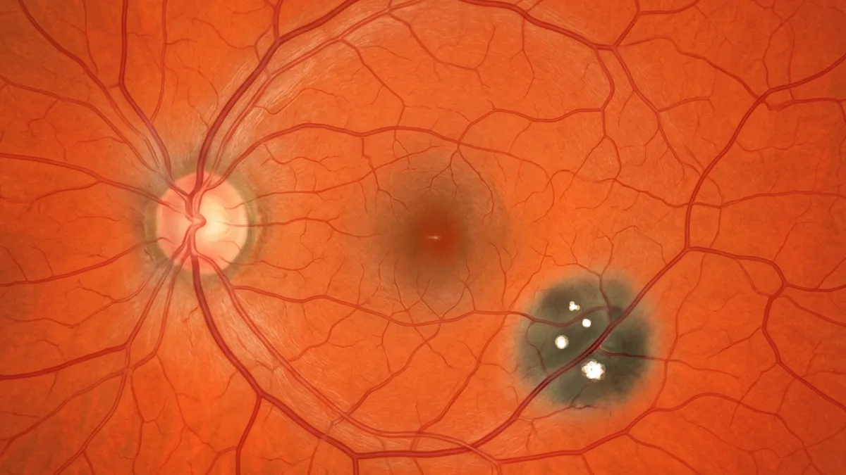

Congenital hypertrophy of the retinal pigment epithelium is a benign and very common lesion of the fundus, present from birth. It appears as a flat, well-defined and heavily pigmented spot, most often discovered by chance. Benign and silent, it mainly needs to be recognised so as not to be confused with a tumour. Dr Julien Gozlan, ophthalmic surgeon specialising in the retina in Paris 16, explains what congenital hypertrophy of the retinal pigment epithelium is, how it is diagnosed and in which cases it warrants further investigation.
What is congenital hypertrophy of the retinal pigment epithelium?
Congenital hypertrophy of the retinal pigment epithelium corresponds to a localised thickening of the retinal pigment epithelium, the thin layer of pigmented cells located beneath the retina. The cells there are larger and more loaded with pigment than normal, which gives the lesion its characteristic dark colour.
It is an abnormality present from birth, of an entirely benign nature. Congenital hypertrophy of the retinal pigment epithelium is not a cancer, virtually never transforms and causes, in the vast majority of cases, no visual disturbance.
What does this fundus lesion look like?
Congenital hypertrophy of the retinal pigment epithelium appears as a flat, round or oval spot, grey-black in colour, with sharp edges. Several features characterise it:
- A flat surface: unlike a tumour, the lesion is not raised.
- Lacunae: small depigmented, lighter areas may appear within it over time.
- A depigmented halo: a thin light rim sometimes surrounds the lesion.
These characteristics help distinguish a congenital hypertrophy of the retinal pigment epithelium from a naevus or a tumour of the choroid.
The different forms
Several presentations of congenital hypertrophy of the retinal pigment epithelium are distinguished:
- The solitary form: a single, isolated lesion, the most frequent and entirely benign.
- The grouped form: several spots clustered in the same sector, suggesting "bear tracks" (animal footprints). This form is also benign.
- The atypical (multifocal) form: small pigmented lesions scattered over both eyes, which may have a particular systemic significance.
What are the symptoms?
In almost all cases, congenital hypertrophy of the retinal pigment epithelium is asymptomatic. The patient feels no discomfort, and the lesion does not affect vision. It is almost always discovered incidentally during a fundus examination carried out for another reason.
Rarely, when it is extensive and affects the centre of the retina, it can be responsible for a small visual field defect (scotoma) corresponding to the pigmented area, without loss of central visual acuity.
How is this lesion diagnosed?
The diagnosis of congenital hypertrophy of the retinal pigment epithelium is based on the fundus examination and imaging performed at Dr Julien Gozlan's practice:
- Fundus photography: it documents the lesion and allows it to be compared over time.
- OCT (optical coherence tomography): it confirms the flat nature of the lesion and the absence of fluid or tumour relief.
- Autofluorescence: it highlights the typical appearance of the hypertrophied pigment epithelium and its lacunae.
These examinations confirm the benign nature of the lesion and rule out a tumour.
Congenital hypertrophy of the retinal pigment epithelium or a pigmented tumour?
The main value of the diagnosis is to differentiate this lesion from a true pigmented tumour:
- The choroidal naevus: deeper, of a more greyish colour, located in the choroid and not in the pigment epithelium.
- The choroidal melanoma: a raised tumour that changes over time, unlike congenital hypertrophy of the retinal pigment epithelium, which remains flat and stable.
The flat, stable and well-defined nature of the lesion is reassuring and strongly points to the diagnosis.
Multifocal form: investigation sometimes needed
The vast majority of congenital hypertrophies of the retinal pigment epithelium, whether solitary or grouped, are associated with no systemic disease and do not increase the risk of cancer.
By contrast, a particular form, made of multiple small "comet-tail" pigmented lesions distributed over both eyes, can be associated with familial digestive polyposis (familial adenomatous polyposis, Gardner syndrome). In this specific case, a digestive work-up and a specialist opinion are recommended. This is why the discovery of bilateral multifocal lesions always deserves careful analysis.
Does it need treatment and monitoring?
Congenital hypertrophy of the retinal pigment epithelium requires no treatment. Management relies on simple monitoring:
- A baseline fundus photograph: to track the stability of the lesion.
- Spaced-out checks: generally every 1 to 2 years, the lesion most often remaining unchanged.
- Appropriate work-up: only in case of a bilateral multifocal form suggesting a systemic cause.
FAQ: congenital hypertrophy of the retinal pigment epithelium
Is congenital hypertrophy of the retinal pigment epithelium dangerous?
No. It is a benign lesion, present since birth, which does not affect vision and virtually never transforms. It simply needs to be recognised and differentiated from a tumour.
Can it turn into a melanoma?
Progression to a malignant tumour is exceptional. Unlike melanoma, congenital hypertrophy of the retinal pigment epithelium remains flat and stable. Spaced-out photographic monitoring is enough to confirm this stability.
Is this lesion linked to colon cancer?
The solitary and grouped forms are not associated with colon cancer. Only the bilateral multifocal form can be a marker of familial digestive polyposis and then warrants an appropriate work-up.
Is treatment needed?
No, no treatment is necessary. Congenital hypertrophy of the retinal pigment epithelium calls for simple monitoring with fundus photography and OCT.
Should my relatives be examined?
For a solitary or grouped form, no family screening is necessary. In the case of a multifocal form suggesting polyposis, a digestive opinion and examination of relatives may be proposed.
When to consult Dr Julien Gozlan?
The discovery of a pigmented spot in the fundus warrants a specialist opinion to determine its nature. Dr Julien Gozlan, retina specialist in Paris 16, has the imaging (fundus photography, OCT, autofluorescence) needed to confirm congenital hypertrophy of the retinal pigment epithelium, distinguish it from a naevus or a melanoma and arrange, if needed, further investigation.
📍 Consultation at the Paris – Auteuil Ophthalmology Practice
Dr Julien Gozlan sees you at the Paris – Auteuil Ophthalmology Practice for the diagnosis and monitoring of congenital hypertrophy of the retinal pigment epithelium. Thanks to a complete retinal imaging platform (fundus photography, OCT, autofluorescence), he precisely characterises your lesion and reassures you.
Book an appointment on DoctolibFurther reading
- Choroidal naevus: discover this benign pigmented lesion of the choroid and its monitoring criteria.
- Choroidal melanoma: understand the main malignant tumour of the fundus and how to distinguish it from a benign lesion.
- Choroidal osteoma: learn about this benign bony tumour of the choroid.
- OCT (optical coherence tomography): a key examination to confirm the flat and benign nature of the lesion.