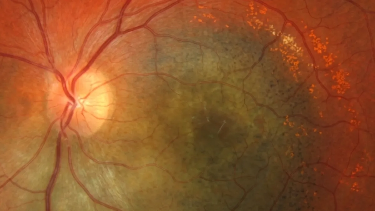

The choroidal melanoma is the most common primary malignant tumour of the eye in adults. Arising from the melanocytes of the choroid — the vascular membrane located beneath the retina — it can remain silent for a long time before affecting vision. Early diagnosis is decisive, as it determines both eye preservation and the patient's prognosis. Dr Julien Gozlan, ophthalmic surgeon specialising in the retina in Paris 16, explains how to recognise a choroidal melanoma, how it is diagnosed and what the current treatment options are.
What is a choroidal melanoma?
The choroidal melanoma is a malignant tumour that develops from melanocytes, the pigment cells present in the choroid. This richly vascularised layer, located between the retina and the sclera, provides nourishment to the photoreceptors. The choroid alone accounts for nearly 90% of uveal melanomas, ahead of the ciliary body and the iris.
With an incidence of about 6 cases per million inhabitants per year, the choroidal melanoma remains a rare tumour. It is nonetheless the most common primary intraocular malignant tumour in adults. It differs from choroidal metastasis, which originates from a cancer located elsewhere in the body.
Choroidal melanoma: which signs should raise concern?
In a large number of cases, the choroidal melanoma is asymptomatic in its early stages and discovered during a routine fundus examination. When it becomes symptomatic, the most frequent signs are:
- Decreased vision: progressive or sudden, especially if the tumour is close to the macula or accompanied by a serous retinal detachment.
- Flashes of light (photopsia): sensations of sparks in the visual field.
- Floaters: appearance of moving dots or filaments in front of the eye.
- Dark spot or visual field defect (scotoma): a fixed area of shadow.
- Image distortion (metamorphopsia): straight lines appear wavy.
These symptoms are not specific to choroidal melanoma, but they always warrant a prompt ophthalmic examination with fundus imaging.
Causes and risk factors of choroidal melanoma
The exact causes of choroidal melanoma are not fully understood. Several factors do, however, increase the risk of developing it:
- Light eyes and skin: people with a fair skin type and blue or green eyes are at higher risk.
- Pre-existing choroidal naevus: a choroidal naevus can, in rare cases, transform into a melanoma.
- Ocular melanocytosis: excessive congenital pigmentation of the eye (oculodermal melanocytosis) increases the risk.
- Genetic predisposition: certain mutations, notably of the BAP1 gene, promote the occurrence of choroidal melanomas.
- Age: the choroidal melanoma is most often diagnosed after the age of 50.
How is a choroidal melanoma diagnosed?
The diagnosis of choroidal melanoma is based on a fundus examination supplemented by multimodal imaging performed at Dr Julien Gozlan's practice:
- Fundus examination: it reveals a pigmented, elevated mass, sometimes mushroom-shaped when the tumour has ruptured Bruch's membrane.
- B-scan ocular ultrasound: it measures the thickness of the tumour and shows choroidal excavation and low internal reflectivity, both highly suggestive of choroidal melanoma.
- OCT (optical coherence tomography): it analyses the retinal impact and the associated subretinal fluid.
- Autofluorescence: it detects orange pigment (lipofuscin), a marker of lesion activity.
- Indocyanine green angiography and fluorescein angiography: they clarify the tumour's vascularisation.
An extension work-up is systematically coordinated with the oncologist, because choroidal melanoma can metastasise, mainly to the liver.
Choroidal melanoma or choroidal naevus: how to tell them apart?
Distinguishing an early choroidal melanoma from a benign naevus is one of the key challenges of the consultation. Ophthalmologists rely on transformation risk factors grouped under the acronym TFSOM-DIM (Shields et al., 2019):
- Thickness: a lesion more than 2 mm thick is suspicious.
- Fluid (subretinal): the presence of fluid is a warning sign.
- Symptoms: decreased vision, photopsia, floaters.
- Orange pigment: visible on autofluorescence.
- Melanoma hollow (hypoechogenicity): a hollow appearance of the lesion on ultrasound.
- DIM (diameter): a diameter greater than 5 mm.
The presence of several of these criteria, or documented growth over time, points to a choroidal melanoma and requires specialised management in ocular oncology.
What treatments are available for choroidal melanoma?
The management of choroidal melanoma is multidisciplinary and aims, whenever possible, to preserve the eye and vision. The choice depends on the size, location and extent of the tumour:
- Brachytherapy (radioactive plaque): placement of an iodine-125 or ruthenium-106 disc against the sclera, the reference conservative treatment for small and medium-sized tumours.
- Proton therapy: high-precision irradiation particularly suited to tumours close to the optic nerve or the macula.
- Transpupillary thermotherapy: laser treatment, often used in combination with irradiation.
- Enucleation: removal of the eye, reserved for large or painful tumours not amenable to conservative treatment.
Regular oncological follow-up always accompanies the local treatment of melanoma in order to detect any distant spread early.
Prognosis and monitoring of choroidal melanoma
The prognosis of the melanoma depends mainly on the size of the tumour, its location and its genetic profile. Small tumours treated early offer a better visual and general prognosis. The main risk is the development of metastases, most often in the liver, sometimes several years after treatment.
Genetic analysis of the tumour (looking for monosomy 3 or a BAP1 mutation) helps assess the metastatic risk. Prolonged, lifelong ophthalmic and oncological follow-up is therefore essential to monitor the treated eye and detect any systemic progression.
FAQ: choroidal melanoma
Is choroidal melanoma a serious cancer?
Choroidal melanoma is a cancer that can threaten vision and, in the event of metastases, the patient's life. Detected early and treated, it is nonetheless well controlled at the ocular level in the majority of cases. This is why early diagnosis and regular follow-up are essential.
Does choroidal melanoma mean losing the eye?
No, not systematically. Conservative treatments such as brachytherapy or proton therapy now make it possible to preserve the eye in most cases. Enucleation is reserved only for very large tumours or those not amenable to conservative treatment.
Can a choroidal naevus become a choroidal melanoma?
This remains rare. The vast majority of choroidal naevi stay benign. It is precisely to detect a possible transformation that regular monitoring of a choroidal naevus is recommended.
Is choroidal melanoma painful?
Most often, no. Choroidal melanoma is generally painless and manifests through visual disturbances. Pain may appear late in the event of secondary glaucoma or a very large tumour.
How is a choroidal melanoma monitored after treatment?
Follow-up combines regular ophthalmic examinations (fundus, OCT, ultrasound) and oncological monitoring with liver imaging. This follow-up is lifelong in order to detect any local recurrence or metastasis as early as possible.
When to consult Dr Julien Gozlan?
Any discovery of an elevated pigmented spot in the fundus, or the appearance of decreased vision, flashes of light, floaters or a visual veil, warrants a prompt specialist opinion. Dr Julien Gozlan, retina specialist in Paris 16, has the multimodal imaging (ultrasound, OCT, autofluorescence, angiographies) needed to confirm or rule out a the tumour and to organise management without delay with an ocular oncology centre.
📍 Consultation at the Paris – Auteuil Ophthalmology Practice
Dr Julien Gozlan sees you at the Paris – Auteuil Ophthalmology Practice for the diagnosis and monitoring of this tumour. Thanks to a complete retinal imaging platform (ultrasound, OCT, autofluorescence, angiography), he provides a precise evaluation of your lesion and coordinates management with ocular oncology.
Book an appointment on DoctolibFurther reading
- Choroidal naevus: discover this benign pigmented lesion and its monitoring criteria.
- Choroidal metastasis: understand this secondary tumour of the choroid and how it differs from melanoma.
- OCT (optical coherence tomography): a key examination for analysing retinal structures and monitoring fundus lesions.
- Indocyanine green angiography: a valuable examination to explore the vascularisation of the choroid.