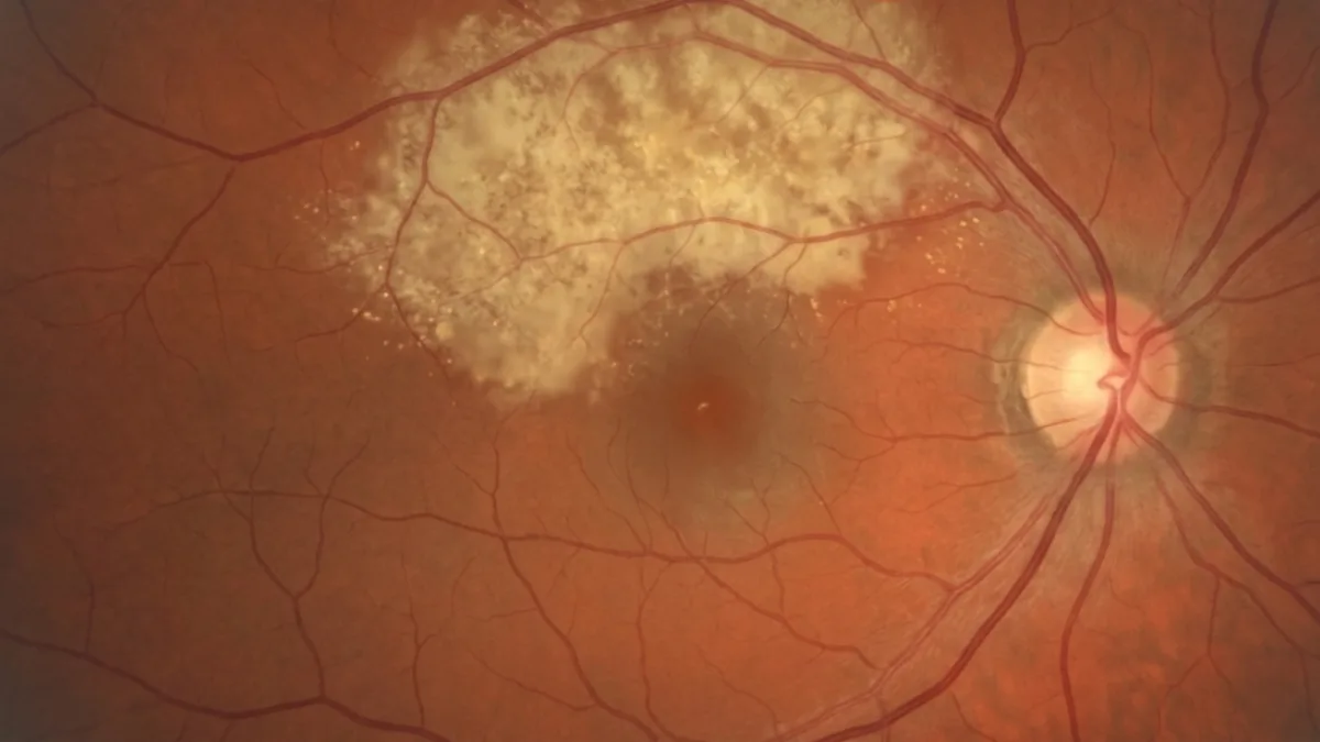

The choroidal osteoma is a rare benign tumour of the choroid, made of mature bone tissue. It mainly affects young women and is most often located near the optic nerve. Although benign, the choroidal osteoma can affect vision when it decalcifies or is complicated by abnormal vessels. Dr Julien Gozlan, ophthalmic surgeon specialising in the retina in Paris 16, explains what a choroidal osteoma is, how it is diagnosed and how it is monitored.
What is a choroidal osteoma?
The choroidal osteoma is a benign bony tumour that develops in the choroid, the vascular membrane located between the retina and the sclera. Surprisingly, it is composed of mature bone, similar to skeletal bone tissue, within a tissue that normally contains none.
It appears as a flat or slightly elevated plaque, yellow-orange in colour, with scalloped edges, most often located near the optic disc. The choroidal osteoma is a rare and entirely benign tumour: it is not a cancer and never gives rise to metastases.
Choroidal osteoma: what are the symptoms?
In many cases, the choroidal osteoma is asymptomatic and discovered incidentally during a fundus examination. When it becomes symptomatic, particularly if it extends towards the macula, it can cause:
- Decreased vision: progressive, related to damage to the photoreceptors over the tumour.
- Image distortion (metamorphopsia): straight lines appear wavy.
- A spot in the visual field (scotoma): corresponding to the lesion.
Sudden vision loss should prompt a search for a complication, in particular the appearance of abnormal vessels under the retina.
Who is affected by choroidal osteoma?
The choroidal osteoma very predominantly affects young women, with a mean age at diagnosis of around twenty years. It is most often unilateral but can sometimes involve both eyes.
The exact cause of the choroidal osteoma remains unknown. It is not linked to trauma or to a deficiency, and it is generally not accompanied by any systemic disease. It is an isolated feature of the fundus.
How is a choroidal osteoma diagnosed?
The diagnosis of the choroidal osteoma is based on the fundus examination and multimodal imaging performed at Dr Julien Gozlan's practice:
- B-scan ocular ultrasound: a key examination, it shows a highly reflective lesion with a characteristic acoustic shadow, the signature of the bony nature of the choroidal osteoma.
- OCT (optical coherence tomography): it analyses the retinal layers above the tumour and detects any fluid or abnormal vessels.
- OCT angiography: it looks for choroidal neovascularisation without injection.
- Autofluorescence and angiography: they clarify the state of the pigment epithelium and the vascularisation of the lesion.
A CT scan can confirm the bony density of the lesion in case of diagnostic doubt.
Choroidal osteoma or another tumour: the differential diagnosis
The yellow-orange appearance of the choroidal osteoma can lead to confusion with other fundus lesions:
- The amelanotic choroidal melanoma: ruled out by ultrasound, which shows no bony acoustic shadow.
- The choroidal haemangioma: of a more pinkish colour and different reflectivity.
- The choroidal metastasis: often associated with a known cancer and with a distinct ultrasound appearance.
The acoustic shadow on ultrasound remains the most specific feature to confirm a osteoma.
Progression and complications of choroidal osteoma
The the osteoma is a benign tumour, but its progression determines the visual prognosis. Two phenomena can impair vision:
- Decalcification: the tumour may lose part of its bone, which weakens the overlying photoreceptors, especially if the fovea is involved.
- Choroidal neovascularisation: the appearance of abnormal vessels under the retina, responsible for bleeding or fluid, is the main cause of severe vision loss.
Overall, a significant proportion of patients with a the tumour may experience vision loss over time, hence the value of regular monitoring.
What treatment for choroidal osteoma?
The tumour itself requires no treatment: a stable the lesion without complications calls for simple monitoring. Management mainly targets complications:
- Intravitreal anti-VEGF injections (IVT): the reference treatment for the choroidal neovascularisation associated with the osteoma.
- Photodynamic therapy: sometimes proposed to treat abnormal vessels.
- OCT monitoring: regular checks to detect decalcification and new vessels early.
FAQ: choroidal osteoma
Is choroidal osteoma a cancer?
No. The the osteoma is a strictly benign tumour, made of mature bone. It does not turn into cancer and never gives rise to metastases. Only its complications can threaten vision.
Can a choroidal osteoma cause vision loss?
Vision loss is possible, especially if the tumour reaches the macula or is complicated by abnormal vessels. Regular follow-up makes it possible to treat these complications early and best preserve vision.
Does a choroidal osteoma need surgery?
No, there is no surgery for the tumour itself. Management relies on monitoring and, if needed, on treating complications with intravitreal injections.
Is choroidal osteoma hereditary?
No, the osteoma is not a hereditary disease and is not accompanied by any systemic condition. It occurs in isolation, most often in young women.
How often should a choroidal osteoma be monitored?
The schedule depends on the location of the lesion and the presence of complications. Monitoring with OCT and fundus examination, generally yearly or more frequent if the macula is threatened, is recommended.
When to consult Dr Julien Gozlan?
Any discovery of a yellow-orange lesion in the fundus, or the appearance of decreased vision, image distortion or a visual spot, warrants a specialist opinion. Dr Julien Gozlan, retina specialist in Paris 16, has the multimodal imaging (ultrasound, OCT, OCT angiography) needed to confirm a the tumour, distinguish it from the other tumours of the choroid and organise its monitoring.
📍 Consultation at the Paris – Auteuil Ophthalmology Practice
Dr Julien Gozlan sees you at the Paris – Auteuil Ophthalmology Practice for the diagnosis and monitoring of a the lesion. Thanks to a complete retinal imaging platform (ultrasound, OCT, OCT angiography), he provides personalised and rigorous follow-up of your lesion.
Book an appointment on DoctolibFurther reading
- Choroidal naevus: discover this benign pigmented lesion of the choroid and its monitoring criteria.
- Choroidal melanoma: understand the main malignant tumour of the choroid and its diagnosis.
- Choroidal haemangioma: learn about this benign vascular tumour of the fundus.
- OCT (optical coherence tomography): a key examination to analyse the retina above the osteoma and monitor its complications.