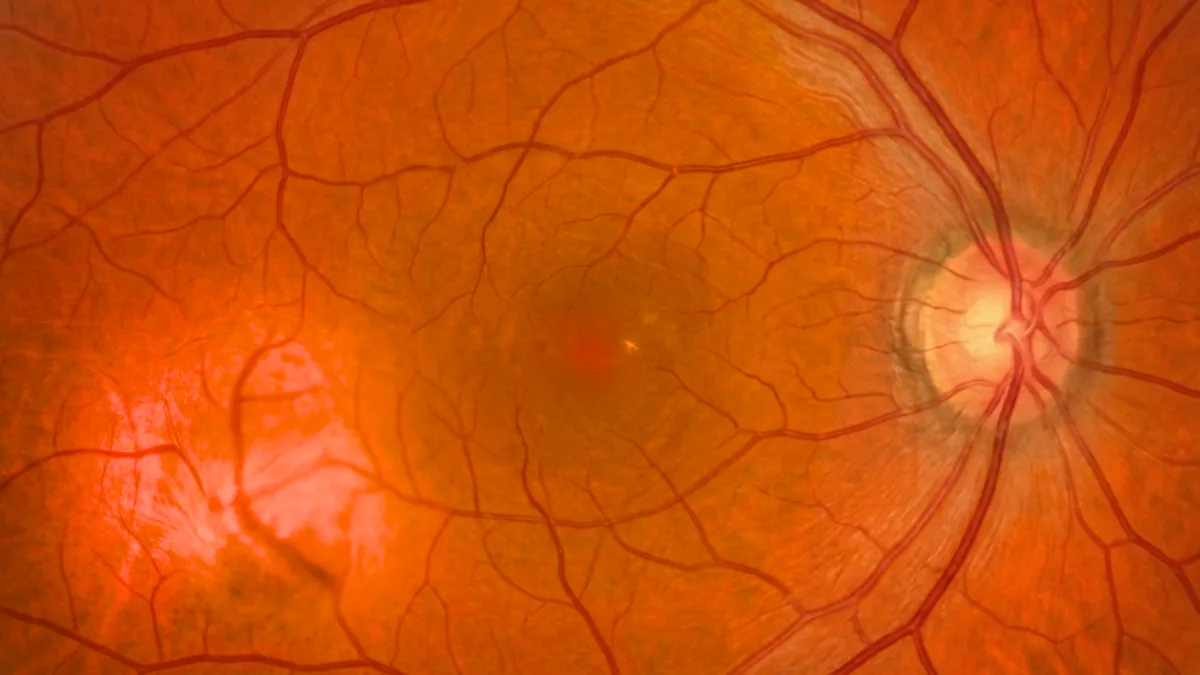

The choroidal haemangioma is a benign vascular tumour of the choroid, that is, a malformation made of normal but abnormally clustered blood vessels. Unlike melanoma, it is not cancerous and never metastasises. It can, however, affect vision when it causes fluid to accumulate beneath the retina. Dr Julien Gozlan, ophthalmic surgeon specialising in the retina in Paris 16, explains what a choroidal haemangioma is, how it is diagnosed and what treatment options are available.
What is a choroidal haemangioma?
The choroidal haemangioma is a vascular hamartoma: a benign lesion made up of dilated blood vessels within the choroid, the richly vascularised membrane located between the retina and the sclera. Clinically, it appears as an orange-pink, elevated mass, most often single and located at the posterior pole of the eye.
This tumour has no potential for malignant transformation. Its visual impact does not depend on cancerous growth, but on the fluid it can leak under the retina, causing a serous detachment and macular oedema.
Circumscribed or diffuse choroidal haemangioma?
Two forms of choroidal haemangioma, very different in presentation, are distinguished:
- The circumscribed form: the most frequent, isolated, with no associated systemic disease. It affects middle-aged adults and remains limited to a well-defined area of the choroid.
- The diffuse form: more extensive, it occurs within the context of Sturge-Weber syndrome (encephalotrigeminal angiomatosis), often associated with a facial cutaneous angioma ("port-wine stain").
This distinction matters, because the diffuse form requires broader management, in connection with the other features of the syndrome.
Choroidal haemangioma: what are the symptoms?
A small choroidal haemangioma can remain completely asymptomatic and be discovered only incidentally. When it becomes symptomatic, it causes:
- Progressive vision loss: especially when the lesion is close to the macula.
- Image distortion (metamorphopsia): straight lines appear wavy.
- A visual field defect: in case of an extensive serous retinal detachment.
- A sensation of veil or spot: linked to the accumulation of subretinal fluid.
These symptoms, related to the subretinal fluid rather than to the tumour itself, warrant a complete fundus examination and retinal imaging.
How is a choroidal haemangioma diagnosed?
The diagnosis is based on the fundus examination and multimodal imaging performed at Dr Julien Gozlan's practice:
- B-scan ocular ultrasound: it shows a mass with high internal reflectivity, a feature that helps distinguish the choroidal haemangioma from melanoma.
- OCT and enhanced depth (EDI) OCT: they analyse the subretinal fluid, macular oedema and the appearance of the choroid.
- Fluorescein angiography: it reveals early vascular filling of the lesion.
- Indocyanine green angiography: a key examination showing early filling followed by late wash-out of the dye, highly suggestive of the diagnosis.
These examinations make it possible to establish the diagnosis and to distinguish the choroidal haemangioma from the other tumours of the choroid.
Choroidal haemangioma or melanoma: how to tell them apart?
This tumour can be confused with a naevus, an amelanotic melanoma or a choroidal metastasis. Several features point to the diagnosis:
- Colour: the choroidal haemangioma is typically orange-pink, whereas melanoma is often pigmented.
- Ultrasound: the high internal reflectivity of the lesion contrasts with the excavation and low reflectivity of melanoma.
- Indocyanine green angiography: the filling and wash-out profile is highly specific.
In case of doubt, comparative monitoring over time and repeated imaging confirm the benign nature of the lesion.
What treatments are available for choroidal haemangioma?
The treatment of choroidal haemangioma depends on the visual disturbance and the presence of subretinal fluid:
- Simple monitoring: an asymptomatic lesion, without fluid, requires no treatment and is followed up regularly.
- Photodynamic therapy (PDT): the reference treatment for symptomatic forms, it leads to regression of the tumour and resorption of the subretinal fluid.
- Transpupillary thermotherapy: laser treatment used in certain locations.
- Radiotherapy (proton therapy, plaque, external beam): reserved for large haemangiomas with extensive retinal detachment.
- Intravitreal anti-VEGF injections: sometimes used as an adjunct to reduce macular oedema.
Prognosis and follow-up of choroidal haemangioma
The prognosis of choroidal haemangioma is generally favourable, since it is a benign tumour that never threatens the patient's life. Preservation of vision depends mainly on the timeliness of treatment when the macula is threatened by subretinal fluid.
Regular ophthalmic follow-up with OCT and fundus examination makes it possible to monitor the lesion's progression, detect any recurrence of fluid and adjust treatment. Early management offers the best chances of preserving good vision.
FAQ: choroidal haemangioma
Is choroidal haemangioma a cancer?
No. It is a strictly benign vascular tumour. It does not turn into cancer and never gives rise to metastases. Treatment may nonetheless be necessary if the lesion affects vision.
Can a choroidal haemangioma cause vision loss?
Vision loss is possible when the tumour causes fluid under the macula. Treated in time, notably with photodynamic therapy, this condition most often regresses and vision can be preserved.
Does a choroidal haemangioma always need treatment?
No. An asymptomatic lesion, without subretinal fluid, only requires monitoring. Treatment is indicated only when there is a visual disturbance or a serous detachment.
What is the difference between a choroidal haemangioma and a melanoma?
The choroidal haemangioma is benign and vascular, whereas melanoma is a cancer. Ultrasound and indocyanine green angiography make it possible to distinguish them with great reliability.
Is choroidal haemangioma hereditary?
The circumscribed form is not hereditary. The diffuse form occurs within Sturge-Weber syndrome, a congenital condition not transmitted in the classic hereditary way.
When to consult Dr Julien Gozlan?
Any vision loss, image distortion or discovery of an orange mass in the fundus warrants a specialist opinion. Dr Julien Gozlan, retina specialist in Paris 16, has the multimodal imaging (ultrasound, OCT, angiographies) needed to confirm a choroidal haemangioma, distinguish it from the other tumours of the choroid and propose the most appropriate treatment.
📍 Consultation at the Paris – Auteuil Ophthalmology Practice
Dr Julien Gozlan sees you at the Paris – Auteuil Ophthalmology Practice for the diagnosis and follow-up of a choroidal haemangioma. Thanks to a complete retinal imaging platform (ultrasound, OCT, angiography), he provides a precise evaluation of your lesion and personalised follow-up.
Book an appointment on DoctolibFurther reading
- Choroidal naevus: discover this benign pigmented lesion of the choroid and its monitoring criteria.
- Choroidal metastasis: understand this secondary tumour of the choroid and its warning signs.
- OCT (optical coherence tomography): a key examination to analyse subretinal fluid and the macula.
- Indocyanine green angiography: a decisive examination for the diagnosis of vascular tumours of the choroid.