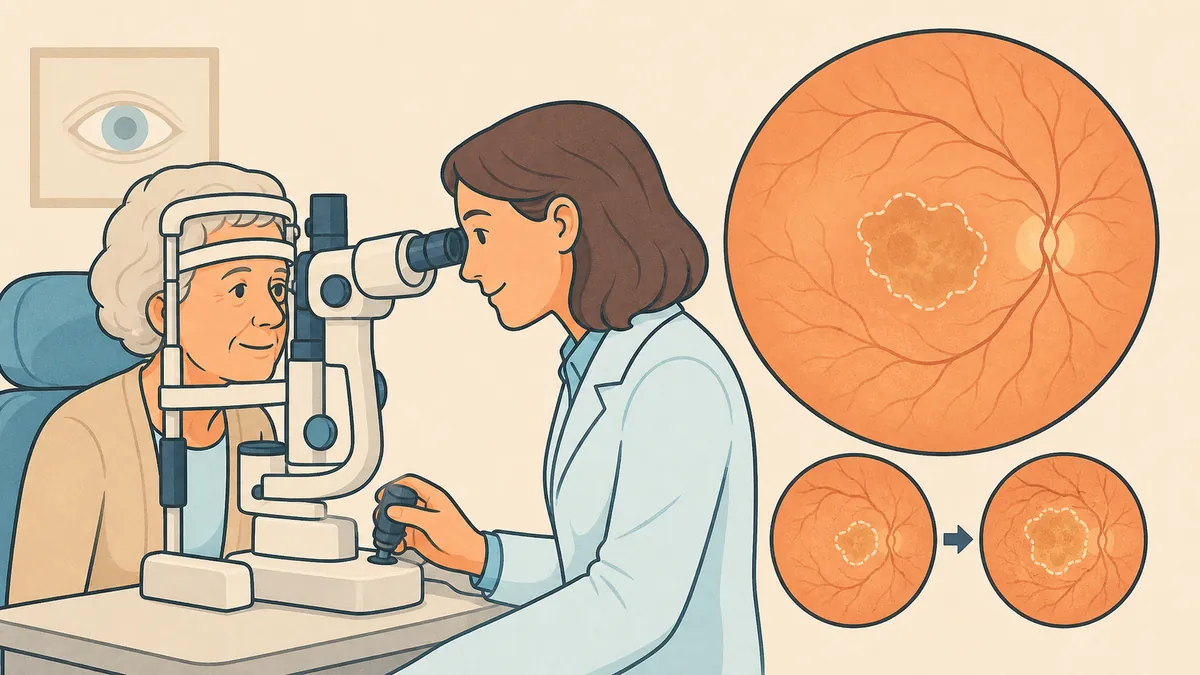

Dry AMD is the most common form of age-related macular degeneration: approximately 8 out of 10 cases. Unlike the wet form, dry AMD progresses slowly, over years, without pain or sudden symptoms. Its advanced stage, called geographic atrophy, corresponds to the gradual disappearance of patches of retina at the center of vision. This article explains how the disease progresses, how to monitor it on a daily basis, and provides an honest assessment — without false hope but without fatalism — of the current state of treatments.
Dry AMD: the most common form of the disease
Age-related macular degeneration exists in two forms. The wet form, caused by the growth of abnormal blood vessels beneath the retina, progresses quickly but can be treated with injections. Dry AMD, also known as atrophic AMD, accounts for approximately 8 out of 10 cases. It affects the macula, the small central area of the retina that enables reading, facial recognition, and the perception of fine detail.
It begins with drusen — small deposits that accumulate beneath the retina — and with changes to the retinal pigment epithelium, the cell layer that nourishes the photoreceptors. At this early stage, vision often remains good and the disease frequently goes unnoticed.
How does dry AMD progress?
Dry AMD progresses slowly, over years. This is a major difference from the wet form, which can cause vision loss within a few weeks. This gradual pace allows time to organize monitoring, adapt your lifestyle, and arrange visual aids if necessary.
The rate of progression varies greatly from one person to another. Some patients retain useful vision for a very long time. Others progress more rapidly, particularly when both eyes are affected or when the lesions approach the center of the macula.
Geographic atrophy: the advanced stage of dry AMD
Geographic atrophy is the advanced stage of the dry form. Well-defined patches of retina, where the visual cells and retinal pigment epithelium have disappeared, become visible on fundus examination. Their outlines resemble a map, hence the term "geographic." These patches gradually expand and eventually reach the center of vision.
As long as the atrophy spares the fixation point, measured visual acuity may remain acceptable, even though reading becomes laborious. When the center is involved, a fixed spot appears in the middle of the visual field.
What symptoms should raise concern?
Dry AMD is painless and progressive. The signs develop silently:
- Difficulty reading: needing more light, missing letters, skipping words at the end of a line;
- Difficulty recognizing faces: details in central vision become unclear;
- Central spot: a blurred, gray, or dark area in the middle of the visual field, at the geographic atrophy stage;
- Distortion of straight lines: this sign is not typical of the dry form and warrants an urgent consultation, as it suggests a wet complication.
How is the diagnosis made?
The diagnosis is based on fundus examination and OCT, a painless, non-contact laser scan of the retina. OCT confirms the diagnosis of dry AMD, defines the extent of the lesions, and measures the expansion of geographic atrophy patches from one visit to the next. OCT angiography can verify the absence of neovascularization without the need for contrast dye injection.
Is there a treatment for dry AMD?
An accurate answer is needed, because the situation differs between continents. In the United States, the FDA approved two drugs injected into the eye in 2023 — pegcetacoplan and avacincaptad pegol. They slow the expansion of atrophy but do not restore vision already lost.
In Europe, no treatment for dry AMD has been approved to date. The European Medicines Agency rejected pegcetacoplan, considering that the benefit perceived by patients had not been sufficiently demonstrated, and the application for the second drug was withdrawn. European patients therefore do not have access to these treatments.
Research remains very active: gene therapies, retinal prostheses — including notable French initiatives. These are clinical trials, not available treatments, and no date for availability can be seriously announced.
Dry AMD: what you can do today
The absence of an approved treatment does not mean the absence of care. Several measures offer real benefit:
- Stop smoking: smoking is the leading modifiable risk factor for the disease;
- Regular monitoring: fundus examination and OCT at intervals agreed with your ophthalmologist;
- Self-monitoring with the Amsler grid: a simple test, one eye at a time, once a week at home;
- AREDS2 supplementation: at the intermediate stage, these supplements reduce the risk of progression to advanced forms;
- Low-vision rehabilitation: magnifying optical aids, adapted lighting, and rehabilitation training to continue reading and maintain independence.
Dry AMD and the risk of wet conversion: staying vigilant
Dry AMD can develop a wet complication at any time — that is, the appearance of neovascularization beneath the retina. This complication can be treated with intravitreal injections, and outcomes are better the earlier treatment is started. Any distortion of straight lines or rapid decline in vision requires an urgent consultation.
FAQ: Dry AMD and geographic atrophy
Does dry AMD cause blindness?
It affects central vision — the vision used for reading and seeing details — but preserves peripheral vision. Dry AMD does not cause total blindness: you can still move around and perceive your surroundings, even at an advanced stage.
Is there a treatment for dry AMD in Europe?
No. Two drugs have been approved in the United States since 2023, but Europe has not approved them: one was rejected by the European Medicines Agency, and the application for the other was withdrawn. Monitoring and preventive measures therefore remain essential.
What is the difference between dry AMD and wet AMD?
The dry form progresses slowly, through the gradual loss of retinal cells. The wet form, caused by abnormal blood vessels, progresses quickly but can be treated with injections. A dry form can convert to a wet form, which is why self-monitoring is so important.
How often should you monitor your vision?
Test each eye separately once a week using the Amsler grid, and follow the consultation schedule set by your ophthalmologist. Any recent change — distortion, spot, or rapid decline — warrants an appointment without waiting for the next check-up.
Are dietary supplements useful?
At the intermediate stage of dry AMD, AREDS2-type supplementation reduces the risk of progression to advanced forms. It does not cure the disease and does not replace quitting smoking or regular monitoring. Talk to your doctor before starting.
When should you see Dr Julien Gozlan?
Dr Julien Gozlan, an ophthalmic surgeon specializing in retinal diseases in Paris 16, provides diagnosis and follow-up for dry AMD: fundus examination, OCT, OCT angiography, and measurement of atrophy progression from one visit to the next. Consult without delay if you notice distorted lines, a recent central spot, or a rapid decline in vision.
📍 Consultation at Cabinet Ophtalmologique Paris - Auteuil
Dr Julien Gozlan welcomes patients at Cabinet Ophtalmologique Paris - Auteuil for a comprehensive macular assessment — fundus examination, OCT, OCT angiography — and regular follow-up of dry AMD.
Book an appointmentFurther reading
- AMD: understanding age-related macular degeneration in both its forms.
- Causes of AMD: age, heredity, smoking — the known risk factors.
- Amsler grid: the self-monitoring test you can do at home.
- AMD and OCT angiography: detecting neovascularization without injection.
- AMD and driving: what the disease changes behind the wheel.