
OCT angiography is a recent imaging technique that allows visualization of retinal blood flow without dye injection. It has become a major tool in the management of age-related macular degeneration (AMD), particularly for detecting and monitoring macular neovascularization. Julien Gozlan, M.D., ophthalmologist in Paris 16, explains the principle of OCT angiography, its specific value in AMD, and how it integrates into treatment monitoring.
What is OCT angiography?
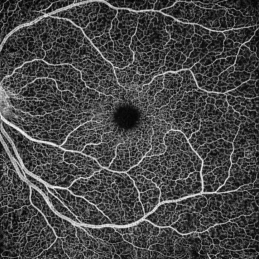
OCT angiography (or OCT-A) is an advancement of macular OCT. While standard OCT shows the structure of retinal layers, OCT angiography analyzes blood flow variations during the examination and reconstructs an image of the vessels, layer by layer.
Unlike fluorescein angiography or indocyanine green angiography, OCT angiography requires neither infusion nor injection. The examination is quick, painless, and can be easily repeated during follow-up.
AMD: why is OCT angiography important?
In the so-called wet (or neovascular) form of AMD, abnormal new vessels develop beneath or within the central retina. These neovessels can leak and cause macular edema, hemorrhages, and distortion of the macula.
OCT angiography allows:
- direct visualization of the neovascular network responsible for wet AMD;
- more precise differentiation of the various forms of neovascularization (type 1, type 2, mixed, etc.);
- assessment of the activity or stabilization of these neovessels during treatment;
- better understanding of certain ambiguous situations on structural OCT or conventional angiography.
How is an OCT angiography examination performed?
The OCT angiography examination is performed on the same device as macular OCT, in a dark room, while seated. You place your chin on a rest, fixate on a light target, and remain still for a few seconds while the device records a series of images.
Practical points to know:
- the examination is painless and requires no injection;
- it takes a few minutes per eye;
- it can be repeated regularly to monitor the progression of AMD;
- good fixation is important to minimize motion artifacts.
Typical OCT angiography findings in AMD
In a patient with neovascular AMD, OCT angiography reveals several characteristic features:
- a choroidal neovascular network visible in the choriocapillaris layer or more superficially;
- a "branching network", "wheel", or "coral" pattern depending on the type of neovascularization;
- hyperflow zones (increased blood flow) corresponding to the core of the neovessel;
- non-perfusion zones or vascular rarefaction around the macula;
- a possible reduction in size or density of the network after effective intravitreal treatments;
- in certain forms, "occult" networks that are poorly visible on fluorescein angiography but clearly identified on OCT angiography.
These findings complement structural OCT (which shows edema, detachments, and atrophy) and allow for a more refined assessment of neovascular activity.
When should OCT angiography be requested in AMD? (decision criteria)
In practice, OCT angiography is particularly useful in the following situations:
- suspected neovascular AMD with edema or serous pigment epithelial detachment on OCT, but inconclusive fluorescein angiography;
- discrepancy between clinical findings, OCT, and conventional angiography (fluorescein / indocyanine green);
- monitoring of a neovessel that appears "nearly dry" on structural OCT to detect possible reperfusion;
- more detailed analysis of polypoidal or mixed forms, in combination with indocyanine green angiography;
- assessment of the treatment plateau (stabilized patients, extended injection intervals) to confirm network inactivity.
The goal is to better tailor the intravitreal treatment strategy and avoid both undertreatment and overtreatment.
OCT angiography, fluorescein angiography, and standard OCT: complementary, not competing
OCT angiography does not entirely replace fluorescein angiography but rather complements it. This can be summarized as follows:
- Structural OCT: shows morphology, edema, detachments, and atrophy;
- Fluorescein / indocyanine green angiography: shows leakage, hemorrhages, and dynamic behavior;
- OCT angiography: directly shows the vascular network without injection, with excellent spatial resolution.
In AMD, the combination of these techniques provides a comprehensive map of the macula and neovascularization, and precisely guides the treatment protocol.
Impact on treatment and intravitreal injection monitoring
The standard treatment for neovascular AMD relies on intravitreal anti-VEGF injections. OCT angiography plays a role at several stages:
- at diagnosis, to confirm the presence of a neovascular network;
- during the loading phase, to analyze the morphological response of the network;
- during the maintenance phase, to help determine whether injection intervals can be extended (treat-and-extend protocols);
- in cases of relapse or visual decline, to distinguish neovascular recurrence from other causes (atrophy, scarring changes).
The goal always remains the same: to preserve vision as much as possible and limit central distortion.
Visual prognosis: what OCT angiography truly provides
OCT angiography is not merely an "impressive" tool for showing vessels. It also provides prognostic information:
- size and density of the neovascular network at diagnosis;
- rate of network reduction under treatment;
- presence of an atrophic zone or non-perfusion around the fovea;
- whether the neovessel is "quiescent" or conversely highly active.
These criteria, analyzed alongside standard OCT and clinical findings, help explain why some patients recover quickly and sustainably, while others require more frequent injections or retain more limited vision.
Practical advice for patients
If an OCT angiography is recommended as part of your AMD management, here are a few simple tips:
- bring your previous imaging if available (past reports, OCT scans, angiographies);
- let your doctor know if you have difficulty staying still or fixating on a target for an extended time;
- understand that the examination carries no systemic risk (no contrast agent is injected);
- do not hesitate to ask questions about the type of AMD, the observed network, and the therapeutic implications;
- strictly follow the monitoring schedule, even if your vision seems stable.
FAQ: AMD and OCT angiography
Is OCT angiography painful?
No. OCT angiography is a painless examination, performed without injection and without contact with the eye in the vast majority of cases. You fixate on a target for a few seconds while the device records the images.
Do I need to fast or prepare anything before the examination?
No, there is no special preparation: you can eat, drink, and take your usual medications. It is simply helpful to bring your previous examinations (OCT scans, angiographies, reports) if available, in order to compare the progression.
Can I drive after an OCT angiography?
If pupil dilation was performed, your vision may be blurry and sensitive to glare for a few hours: it is then advisable to avoid driving right after. Without dilation, driving is generally possible if your vision is comfortable.
How is OCT angiography useful in AMD?
In neovascular AMD (wet form), OCT angiography allows direct visualization of the neovascular network beneath or within the retina, without injection. It helps confirm the presence of neovascularization, specify its type (for example type 1, type 2, mixed), and monitor its appearance over time, in addition to structural OCT, which shows edema, detachments, and atrophy.
Can OCT angiography replace fluorescein or indocyanine green angiography?
No, not entirely. OCT angiography primarily shows the vascular network (flow), but it does not directly visualize leakage like fluorescein angiography, and it is less effective at exploring the retinal periphery. In AMD, the examinations are often complementary: structural OCT for activity (fluid), angiography for leakage dynamics, and OCT-A for detailed mapping of the network.
Why are artifacts mentioned in OCT angiography?
Because OCT-A reconstructs vessels from micro-variations in signal related to blood movement. Eye movements, unstable fixation, cataract, opacities, or segmentation errors can create false images (or mask actual flow). This is why the analysis must always verify acquisition quality and be cross-referenced with structural OCT.
Is OCT angiography sufficient to determine whether AMD is "active"?
Not on its own. The activity of neovascular AMD is primarily assessed on structural OCT (presence of intraretinal or subretinal fluid, detachments, hemorrhages) and on clinical examination. OCT angiography provides additional information: it shows the presence and architecture of the network, and can help identify a "quiescent" neovessel or revascularization, but it does not replace fluid analysis on OCT.
At what point is it requested during injection (anti-VEGF) follow-up?
It is useful at diagnosis to confirm a neovascular network, then at certain key points during follow-up: in cases of discrepancy between symptoms, OCT, and angiography, when there is doubt about reactivation, or when one wishes to document a network in a "nearly dry" context before extending the injection intervals (treat-and-extend protocols). The frequency of repetition therefore depends on the clinical situation, not on a fixed schedule.
How long does the examination take?
A few minutes. The acquisition itself takes a few seconds per eye, but several scan fields (for example 3×3, 6×6, 12×12) may be performed depending on what needs to be analyzed, and a repeat scan may be necessary if fixation was not optimal.
What should I monitor at home if I am being followed for AMD?
Monitor your vision one eye at a time, particularly reading and straight lines (Amsler grid if recommended to you). Any new distortion, central vision loss, appearance of a spot in the center, or unusual discomfort should prompt you to contact the practice promptly, even if a follow-up appointment is already scheduled. OCT angiography is a monitoring tool, but responsiveness to symptoms remains essential.
When to consult Julien Gozlan, M.D.?
You can consult Julien Gozlan, M.D. if you have AMD, suspected macular neovascularization, or if intravitreal injection monitoring has been recommended to you. A comprehensive assessment combining OCT, OCT angiography, and clinical examination allows for an accurate diagnosis and the definition of a personalized treatment strategy.
The goal is to stabilize your vision as effectively as possible, limit central distortion, and adjust the frequency of injections to the actual disease activity.
📍 Consultation at Paris – Auteuil Ophthalmology Practice
Julien Gozlan, M.D. sees you at Paris – Auteuil Ophthalmology Practice for a comprehensive AMD assessment, including OCT, OCT angiography, and a detailed discussion of intravitreal treatment options.
Book an AppointmentFurther reading
- Age-related macular degeneration (AMD): dry and neovascular forms.
- Intravitreal injections: procedure, efficacy, and follow-up.
- Macular OCT: the structural examination complementary to OCT angiography.
- Intravitreal injections: principle, procedure, and follow-up.